

Autophagy is a fundamental cellular process that helps eliminate damaged components and invading pathogens. But new research published in Nature Immunology reveals that autophagy can also act as a brake on protective innate immune responses in the lungs.
Researchers found that reducing autophagy in alveolar macrophages (AMs) enhanced host resistance against both Staphylococcus aureus and influenza A virus (IAV) in preclinical models (Figure 1).
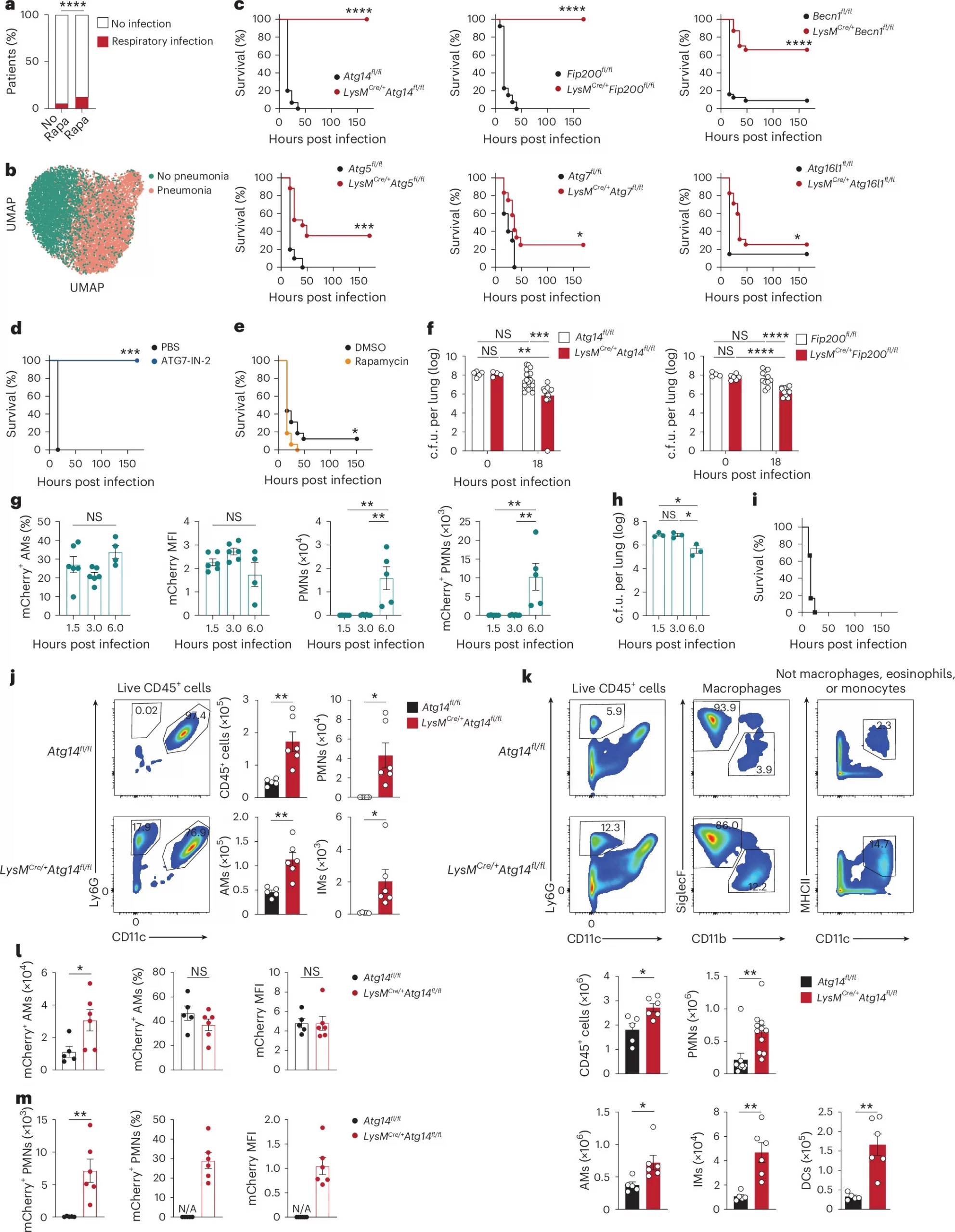
Figure 1: Autophagy deficiency enhances clearance of pulmonary S. aureus. a, Percentage of patients with and without a diagnosis of respiratory infection who were treated with rapamycin (Sirolimus) (n = 1,396 (1,004 females), median age 57 years (range, 45–66 years)) or not (n = 16,436 (11,078 females), median age 56 years (range, 43–66 years)) in the rheumatic patients cohort. P values were determined by chi-square test. b, Dimensionality reduction analysis in individuals with or without bacterial pneumonia15 using an autophagy gene set. Dot color represents different disease groups. c, Survival of Becn1fl/fl (n = 28), LysMCre/+Becn1fl/fl (n = 23), Atg5fl/fl (n = 10), LysMCre/+Atg5fl/fl (n = 17), Atg7fl/fl (n = 10), LysMCre/+Atg7fl/fl (n = 12); Atg16l1fl/fl (n = 8), LysMCre/+Atg16l1fl/fl (n = 17), Atg14fl/fl (n = 15), LysMCre/+Atg14fl/fl (n = 15), Fip200fl/fl (n = 13) or LysMCre/+Fip200fl/fl (n = 10) mice at 0 to 168 h after intranasal (i.n.) inoculation with 4 × 10⁸ colony-forming units (c.f.u.) S. aureus strain USA300. Data are pooled from three or four experiments, P by log-rank Mantel–Cox test. d, Survival of wild-type C56BL/6 J mice treated intratracheally (i.t.) with ATG7-IN-2 (3 mg kg−1 daily for 8 days) (n = 6) or PBS (n = 7) before i.n. infection of S. aureus. Data are pooled from two experiments. P by log-rank Mantel–Cox test. e, Survival of wild-type mice treated intraperitoneally (i.p.) with rapamycin (1.5 mg kg−1 for 6 h) (n = 16) or DMSO (n = 16) before i.n. inoculation of S. aureus. Data are pooled from three experiments. P by log-rank Mantel–Cox test. f, Quantification of S. aureus c.f.u. in the right lung of mice at 0 h (Atg14fl/fl, n = 5; LysMCre/+Atg14fl/fl, n = 4; Fip200fl/fl, n = 4; LysMCre/+Fip200fl/fl, n = 6) and 18 h (Atg14fl/fl, n = 17; LysMCre/+Atg14fl/fl, n = 16; Fip200fl/fl, n = 11; LysMCre/+Fip200fl/fl, n = 11) after i.n. infection. Data are pooled from two or three experiments; mean ± standard error of the mean (s.e.m.). Each dot represents a mouse. Adjusted P values were calculated by Sidak’s multiple comparisons test. g, Percentage of mCherry+SiglecF+ AMs, mCherry mean fluorescence intensity (MFI) in mCherry+ AMs, and number of Ly6G+ neutrophils (PMNs) and mCherry+ PMNs in BAL of wild-type mice (n = 4–5) infected with 4 × 10⁸ c.f.u. mCherry-USA300. Data are pooled from two experiments, mean ± s.e.m., each dot represents a mouse. P by unpaired two-tailed t test. h, S. aureus c.f.u. in the right lung of wild-type mice (n = 3) after i.n. infection with mCherry-USA300. Representative of two experiments, mean ± s.e.m., P by unpaired two-tailed t test. i, Survival of wild-type mice (n = 5) after i.n. inoculation with S. aureus. P by log-rank Mantel–Cox test. j, Representative flow cytometry plots and quantification of CD45+ immune cells including SiglecF+ AMs, Ly6G+ PMNs and CD11b+ interstitial macrophages (IMs) in the BAL of naive Atg14fl/fl (n = 5) or LysMCre/+Atg14fl/fl (n = 6) mice. Data are pooled from two experiments, mean ± s.e.m. P by unpaired two-tailed t test. k, Representative flow cytometry (top) and quantification (bottom) of PMNs (Atg14fl/fl, n = 9; LysMCre/+Atg14fl/fl, n = 11), AMs, IMs, and DCs (Atg14fl/f, n = 5; LysMCre/+Atg14fl/fl, n = 6) in the lungs of naive uninfected mice. Data pooled from two or three experiments, mean ± s.e.m. P by unpaired two-tailed t test. l,m, Number and percentage of mCherry+ AM and mCherry MFI in AMs (l) or number and percentage of mCherry+ PMNs and mCherry MFI in PMNs (m) in the BAL of Atg14fl/fl (n = 5) or LysMCre/+Atg14fl/fl (n = 6) mice at 1.5 h after i.n. inoculation with mCherry-USA300. Data are pooled from two experiments, mean ± s.e.m. P by unpaired two-tailed t test. N/A, not available; NS, not significant.
Under normal conditions, basal autophagy suppressed the expression of interleukin-1α (IL-1α) in alveolar macrophages, limiting innate immune priming.
When autophagy genes were inactivated in these cells, IL-1α expression increased, triggering stronger IL-1R–MyD88 signalling in neutrophils and promoting the recruitment of additional myeloid cells into the lung.
The result? Autophagy-deficient mice cleared S. aureus more efficiently, with reduced lung damage and inflammation.
The researchers identified an immune regulatory pathway linking tissue-resident macrophages to broader myeloid responses:
- Alveolar macrophages: Reduced autophagy releases the restraint on IL-1α expression.
- Neutrophils: Increased IL-1R–MyD88 signalling amplifies innate immune responses.
- Lung tissue: Enhanced myeloid cell recruitment improves pathogen control.
Notably, overexpressing IL-1α specifically in alveolar macrophages also conferred protection against both S. aureus and IAV.
This protection was independent of the microbiome and adaptive immunity, and relied on IL-1α signalling.
These findings highlight IL-1α-mediated innate immune priming as a potential strategy for broad-spectrum protection against respiratory infections, including antibiotic-resistant bacterial pathogens.
However, enhancing inflammation is a delicate balance. Although the mice showed improved bacterial clearance alongside reduced lung damage, translating this approach into humans will require careful investigation of safety, timing and immune regulation.
This study reveals that autophagy does more than support cellular housekeeping and pathogen degradation. It also helps determine the baseline immune readiness of tissue-resident macrophages.
Understanding how to fine-tune this balance could open new avenues for host-directed therapies that strengthen resistance to diverse respiratory pathogens.
Journal article: Cui, X., et al. 2026. Autophagy in macrophages curtails IL-1α-mediated immune priming against lung infections. Nature Immunology.
Summary by Stefan Botha