


CD1a is highly expressed on Langerhans cells, where it presents lipid antigens to T cells. But its in vivo function has been difficult to study because mice naturally lack CD1a.
Researchers engineered mice to express human CD1a and uncovered a surprising role in inflammation (Figure 1).
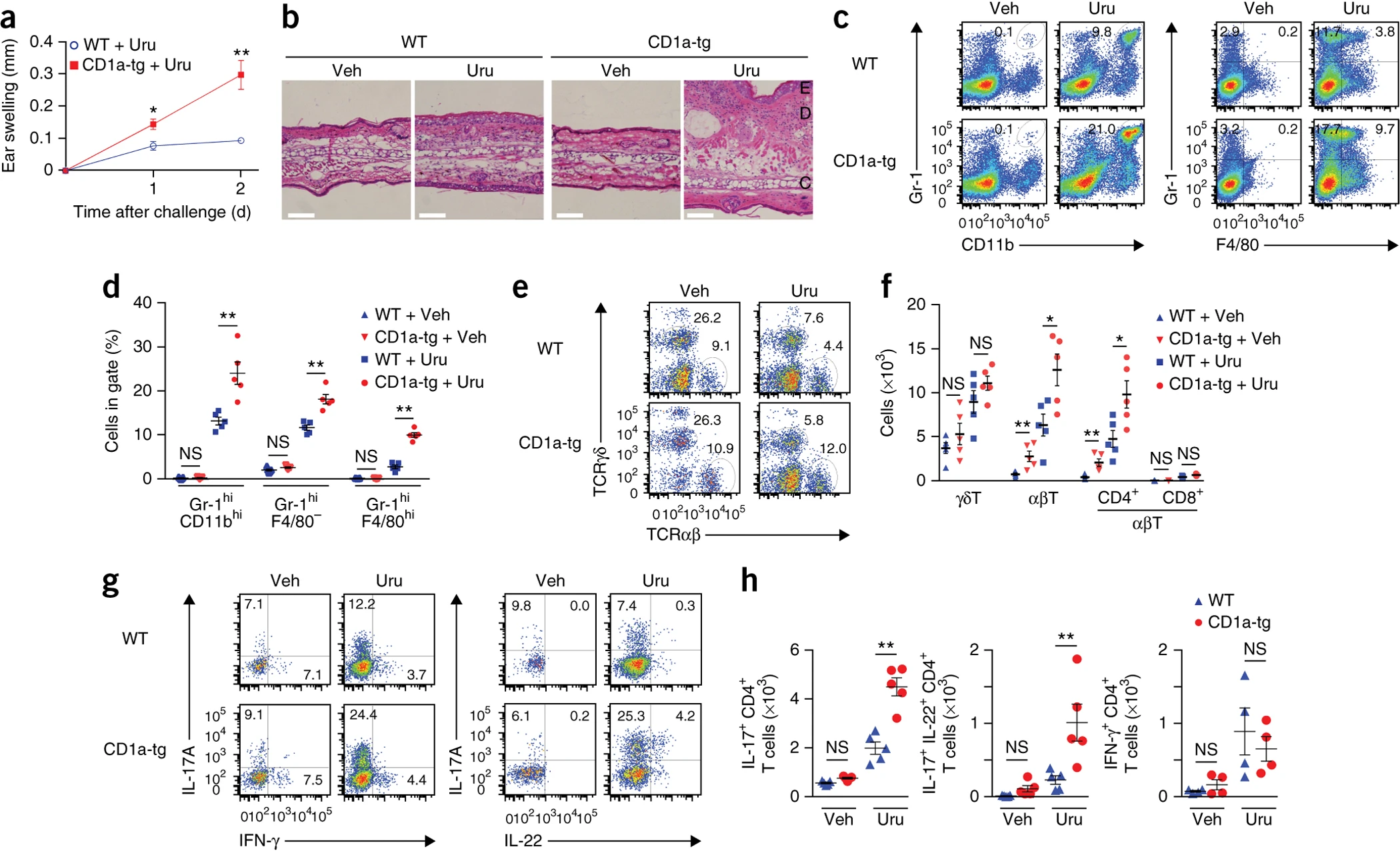
Figure 1: CD1a facilitates TH17 cell–mediated skin inflammation caused by poison ivy. (a) Ear swelling in wild-type (WT) and CD1a-tg mice (n = 5 per group) sensitized by painting of urushiol on the abdomen on day 0 and challenged with urushiol (Uru) or vehicle (Veh; not shown in a) on the ear on day 5, assessed on day 2 after challenge. (b) Microscopy of cross-sections of ears from mice as in a, stained with hematoxylin and eosin. C, cartilage; D, dermis; E, epidermis. Scale bars, 100 μm. (c–h) Flow cytometry (c,e,g) of granulocytes, macrophages and T cell subsets in ear skin of mice as in a, assessed 2 d after challenge, with quantification of results as frequency (d) or absolute number (f,h). (c) Numbers adjacent to outlined areas (left) indicate percent Gr-1hiCD11bhi inflammatory granulocytes among all live cells; numbers in quadrants (right) indicate percent F4/80+Gr-1+ macrophages (top right) or F4/80−Gr-1+ granulocytes (top left) among all live cells. (e) Numbers adjacent to outlined areas indicate percent αβ T cells (bottom right) or γδ T cells (top left) among live CD45+ cells. (f) Absolute number of cells in various T cell subsets (horizontal axis). (g) Numbers in quadrants indicate percent IL-17A+IFN-γ− cells (top left) or IL-17A−IFN-γ+ cells (bottom right) (left), or IL-17A+IL-22− cells (top left) or IL-17A+IL-22+ cells (top right) (right), among TCRβ+CD4+ cells. (h) Absolute number of cytokine-producing T cell subsets as in g. Each symbol (d,f,h) represents an individual mouse; small horizontal lines indicate the mean (± s.e.m.). NS, not significant (P > 0.05); *P < 0.05 and **P < 0.01 (unpaired t test (a) or Mann-Whitney test (d,f,h)). Data are from one experiment representative of five independent experiments with similar results (mean ± s.e.m. in a).
Exposure to urushiol, the plant-derived lipid responsible for poison ivy dermatitis, triggered CD1a-dependent skin inflammation driven by TH17 cells producing IL-17 and IL-22.
One urushiol component, C15:2, was identified as the dominant antigen recognised by CD1a-restricted T cells. Structural analysis then revealed how this lipid fits into CD1a’s antigen-binding cleft.
Importantly, people with poison-ivy dermatitis showed a similar IL-17/IL-22 inflammatory signature.
The researchers also found that CD1a amplified TH17 responses to self-lipids in psoriasis. Blocking CD1a reduced skin inflammation in experimental models.
The bigger picture: CD1a may act as an important bridge between lipid recognition and chronic skin inflammation, making it a potential therapeutic target in diseases such as psoriasis.
Journal article: Kim, J.H, et al. 2016. CD1a on Langerhans cells controls inflammatory skin disease. Nature Immunology.
Summary by Stefan Botha