


In a recent study, researchers have unveiled the immune cell landscape of a healthy human cornea, revolutionizing our understanding of its protective mechanisms against pathogens and diseases (Figure 1).
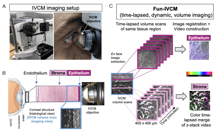
Figure 1: Overview of the Fun- IVCM method. (A) Laser- scanning, IVCM setup with the Heidelberg HRT- 3 with the Rostock Corneal Module, to acquire images of the human cornea (B), shown diagrammatically in histological cross- section [not to scale]. (C) The Functional IVCM (Fun- IVCM) method involves sequentially capturing high- resolution en face volume (z- stack) images [400(x) × 400(y) × 100(z) μm], spanning the basal layer of the corneal epithelium to the mid- stroma. Repeat volume scans of the same region are acquired every 4 to 7 min for a total of 25 to 40 min. En face images at the same tissue plane are extracted from the volume scans, registered to stationary landmarks, and time- lapsed videos are reconstructed at both the level of the corneal epithelium and anterior stroma.
By employing a newly developed imaging technique called Functional In Vivo Confocal Microscopy (Fun-IVCM) alongside advanced analytical approaches, the study revealed that a substantial number of cells present on the surface of the healthy cornea are T cells.
The researchers observed these T cells to be highly dynamic, swiftly moving around and engaging with other cells and nerves in the outermost layer of the cornea. Moreover, they documented distinct cell behaviors in response to factors such as contact lens wear and allergic eye disease. They also quantified how these behaviors could be influenced by drug treatments.
These findings fundamentally reshape our knowledge of the various immune cell subsets in the human cornea and their responses to different stimuli. The discovery of changes in T cells and their behaviors serves as a crucial clinical biomarker of diseases and holds promise in guiding more effective treatments.
Journal article: Downie, L. E., et al., 2023. Redefining the human corneal immune compartment using dynamic intravital imaging. PNAS.
Summary by Stefan Botha


