


Cancer immunotherapy has revolutionised the treatment landscape for various types of cancer by harnessing the power of the immune system to combat tumours. Specifically, immunotherapies that inhibit checkpoint receptors like PD-1, which curtail the ability of T cells to target cancer cells, have emerged as preferred options for treating solid cancers.
The use of PD-1-blocking agents can lead to unintended consequences, as T cells may attack not only cancer cells but also healthy tissues. This phenomenon results in severe and potentially life-threatening side effects, which can diminish the benefits of immunotherapy.
A recent study has provided valuable insights into the underlying mechanisms through which PD-1 maintains the integrity of healthy tissues (Figure 1). These findings have the potential to aid scientists in predicting, treating, or even preventing the side effects associated with PD-1-blocking immunotherapies.
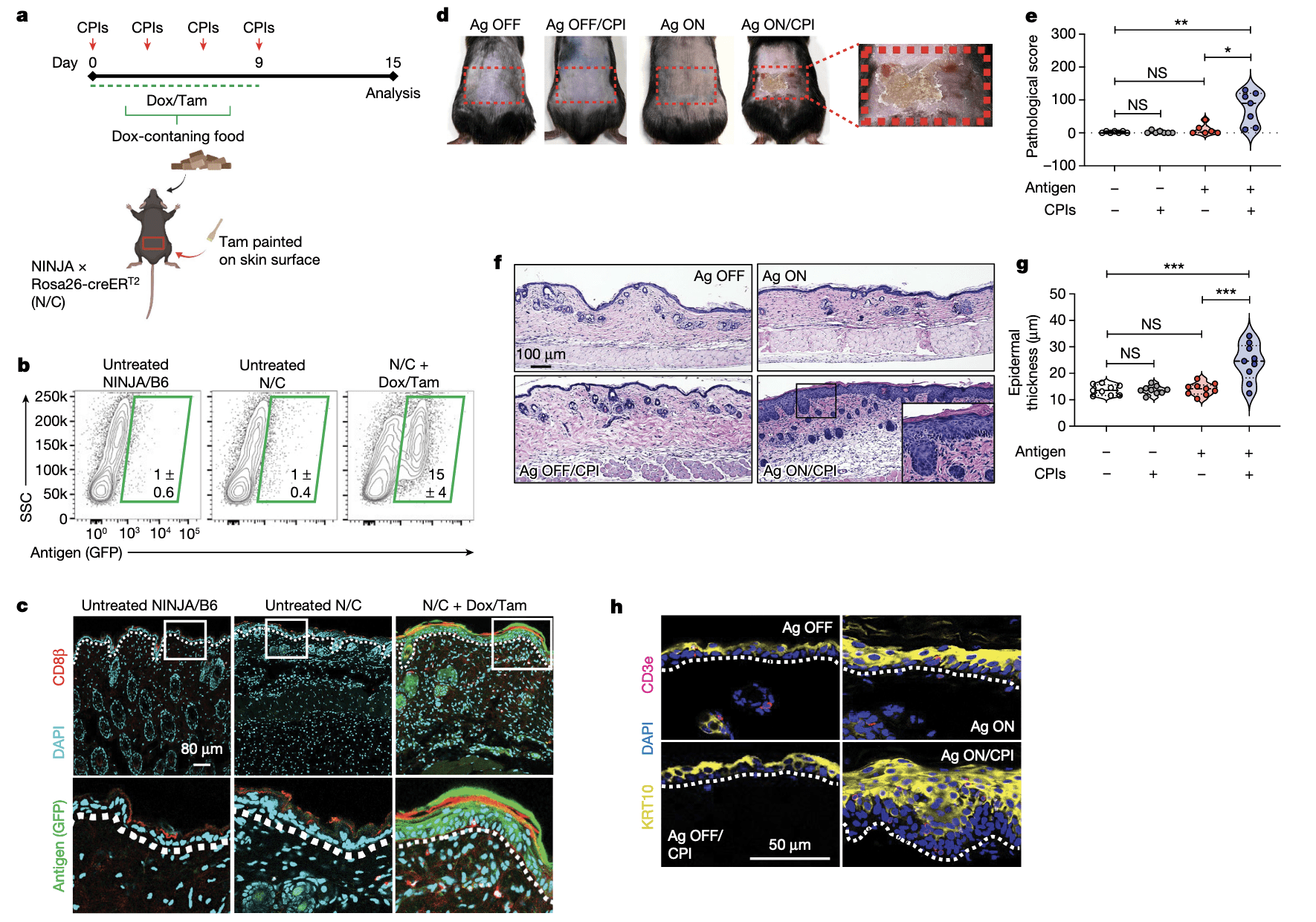
Figure 1: Skin-specific antigen induction and CPIs lead to local cutaneous disease. a, Experimental schedule. b, Frequencies of antigen-expressing (GFP +) skin cells from the indicated conditions (outlined in green; percentages of GFP + cells are shown). Data are mean ± s.d. n = 5 (untreated NINJA, B6 and N/C) or 7 (N/C + Dox/Tam), representative of 4 experimental repeats. c, Confocal microscopy of skin from the indicated conditions. The dotted line indicates the epidermis (top)–dermis (bottom) interface. The outlined region is magnified in the bottom row. n = 3, representative of 2 experimental repeats. d, Representative images of the back of experimental mice treated as indicated. The red rectangle indicates the Tam-treated area that expresses the NINJA antigens. n = 6 (Ag ON) or 7 (Ag OFF, Ag OFF/CPI and Ag ON/CPI), representative of 3 experimental repeats. e, Pathological scores by experimental condition. * P = 0.0112, ** P = 0.0023; NS, not significant by two-tailed t-test. n = 6 (Ag ON) or 7 (Ag OFF, Ag OFF/CPI and Ag ON/CPI), representative of 3 experimental repeats. f, Haematoxylin and eosin (H&E) staining of skin sections from the indicated conditions. Inset shows an expanded view of the outlined area. n = 6 (Ag ON) or 7 (Ag OFF, Ag OFF/CPI and Ag ON/CPI), representative of 3 experimental repeats. g, Quantification of epidermal thickness under indicated conditions. *** P = 0.0006 (Ag ON/CPI versus Ag OFF), *** P = 0.0008 (Ag ON/CPI versus Ag ON); NS, not significant by two-tailed t-test. n = 3 ROIs per mouse for 3 mice, representative of 3 experimental repeats. h, Confocal microscopy of skin from the indicated conditions. Dotted line shows the epidermis (top)–dermis (bottom) interface. n = 5.
For the study, researchers employed innovative mouse models designed to elucidate the role of PD-1 in preventing T cells from attacking healthy skin. By blocking PD-1 and mimicking the effects of immunotherapy, the researchers observed the development of skin disorders in the mice, like those observed in cancer patients receiving PD-1 inhibitors.
These research findings lay the foundation for the future development of enhanced immunotherapies that can mitigate or prevent adverse events. By gaining a better understanding of the vital role played by PD-1 in safeguarding normal tissues from T cell-mediated damage, scientists can strive towards devising strategies that maximize the therapeutic benefits of PD-1 blockade while minimizing harmful side effects.
Journal article: Damo, M., et al., 2023. PD-1 maintains CD8 T cell tolerance towards cutaneous neoantigens. Nature.
Summary by Stefan Botha


