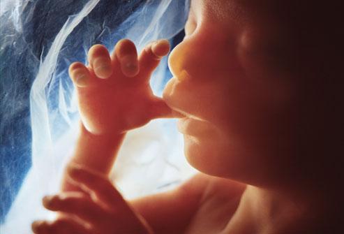
Development at 20 Weeks
The baby weighs about 10 ounces and is a little more than 6 inches long. Your uterus should be at the level of your belly button. The baby can suck a thumb, yawn, stretch, and make faces. Soon — if you haven’t already — you’ll feel your baby move, which is called “quickening.” Source: WebMD
A foetus is a semi-conceptus which survives the maternal immune attack leading to the maintenance of pregnancy to full term. A galore of immune cells are present at the feto- maternal interface and the modulation of immune response at this interface aids in the survival of semi-allogenic foetus. The transmembrane protein, Programmed cell death protein 1 (PD-1) is expressed by a variety of activated immune cells, including T (CD4+, CD8+, NKT-like and regulatory T (Treg)) cells, B cells, monocytes and dendritic cells. Extensive research has shown PD-1 to be involved in T cell homeostasis and along with its ligands helps in the maintenance of peripheral immune tolerance. What is less known is the importance of PD-1/PDL1 pathway in maternal-foetal immunotolerance.
Meggyes et.al in this paper has highlighted the role of PD-1/PDL1 pathway during pregnancy. They recruited healthy women who underwent pregnancy termination for non-medical reasons in the first trimester of their pregnancy. The control group consisted of healthy non-pregnant women. Results revealed an increase in the proportion of decidual immune cells subsets, CD8+ T-, CD56 + NK-, CD56dimNK, and CD56brightNK cells from the decidual tissue compared to the peripheral blood. Decidual CD8+ T, CD4+ T, and NKT-like cells showed an increased expression of PD-1 compared to the periphery. Authors propose that the increase in the expression of PD-1 receptor by the residual CD8+ cells would be to regulate and avoid exaggerated inflammatory effects during pregnancy.
Apart from the expression of PD-1 on the decidual immune cells, authors also analysed the cytotoxic potential of these cells. Results showed a significantly decreased cytotoxic activity by decidual PD-1+ CD8+ T cells compared to the peripheral counterparts. NKG2D, an activation receptor and its co-expression with PD-1 was analysed and it was seen that NKG2D expression was decreased by the PD-1+ CD8+ T subsets in the first trimester compared to non- pregnant condition but the expression level of the decidual counterparts was significantly elevated compared to the periphery. There was decreased cytotoxic activity via decreased CD107a expression by decidual PD-1/NKG2D double positive CD8+ T cells. Authors do suggest that PD-1+ NKG2Dhigh CD8+ T cell interaction via surface NKG2D ligands by the trophoblast could help in the invasion of extravillous trophoblast and may also benefit in preventing microbial infections. Thus, PD-1/PDL1 pathway aids in the maintenance of the immunological environment at the foetal-maternal interface.
Journal article: Meggyes et.al, 2019. The importance of the PD-1/PD-L1 pathway at the maternal-fetal interface. BMC Pregnancy and Childbirth
Article by Babita Balakrishnan