


Researchers have developed a completely new theory about how immune cells identify dangers like viruses in a recent study. The finding may help scientists create better vaccines and acquire a better understanding of autoimmune conditions and allergies (Figure 1).
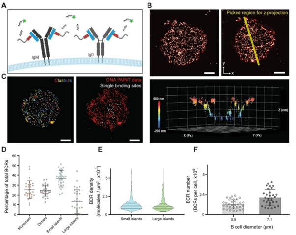
Figure 1: B cell receptor distribution of resting, naïve murine B cells. A An anti-mouse kappa light chain Nb (κLC-Nb) which binds both IgM and IgD BCRs, conjugated site-specifically to a single docking strand, was used for imaging by the DNA-PAINT method (kappaLc: blue; Nb: red; DNA: black; Cy3b: green). B Both 2D (top left panel) and 3D (bottom panel) images of the BCRs were acquired. Plotting a rectangular region in xyz (top right panel) shows a preservation of the round architecture of B cells (bottom panel). Px, pixels. C To extract the information on the number of BCRs on the B cell surface along with the surface area, representative regions of the imaged B cells were analyzed by DBSCAN cluster analysis to identify clusters (left panel and Supplementary Fig. 4). For kinetic calibration, single binding sites were picked (right panel, and Supplementary Fig. 2). Imaging data are representative of 3 independent experiments. D After calculating the number of BCRs per cluster detected by DBSCAN, the percentage of BCRs found as monomers (1 molecule), dimers (2 molecules), small islands (3–9 molecules) and large islands (>9 molecules) was determined. Bars indicate mean ± SD of a total of n = 31 cells from two independent experiments. E qPAINT analysis was used to calculate the BCR density on the imaged surface, taking into consideration the labeling efficiency (Supplementary Fig. 3). The lines in the violin plots indicate the quartiles and the median. F Total BCR numbers were estimated to be around 15,000 for 5.5 μm B cell diameter (literature27) and 25,000 for 7.1 μm diameter (measured). Bars indicate mean ± SD. For images, all scale bars are 1 μm.
In the study, experts share important new information about B cells, which are an essential component of the body’s defensive system. When we are immunised or infected, B cells are the cells that produce protective antibodies; however, B cells are also the cells that create damaging antibodies in association with allergies or autoimmune illnesses. The activation mechanism that is initiated when the cells identify a particular target or “enemy”—an antigen—was investigated by the researchers as the first stage in activating the B cells.
Before, it was thought that antigens from, say, viruses or immunizations, would need to cross-bind a B-cell’s cell surface receptors. That is what all of the texts state. They have demonstrated that B cells can be activated by proteins that can only engage one receptor at a time.
Because the researchers have revealed new information regarding the basis for how receptors on the surface of cells transmit messages into the cells — a crucial biological process — the finding is intriguing for the immunological field as well as for cell biology in general.
They might have disproven the thirty or forty-year-old theory that described how B cell stimulation occurs.
Journal article: Ferapontov, A., et al., 2023. Antigen footprint governs activation of the B cell receptor. Nature Communications.
Summary by Stefan Botha


