


In a recent study conducted in mice, researchers have gained new insights into understanding the mechanisms underlying brain damage in progressive multiple sclerosis (MS) (Figure 1). The study focused on inflammation in the brain’s protective barrier, known as the meninges, and its potential spill-over effects into the adjacent grey matter, which could contribute to the progression of the autoimmune disease.
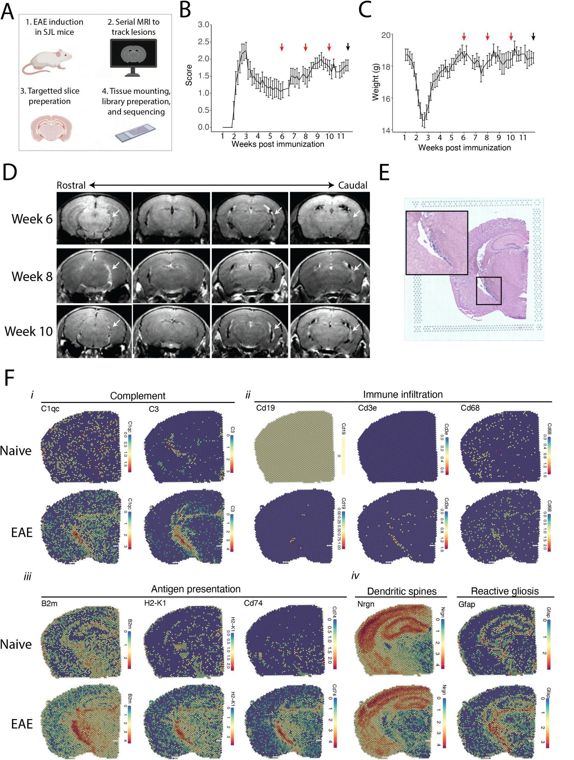
Figure 1: MRI guided spatial transcriptomics of meningeal-based inflammation in SJL EAE. (A) Schematic describing the experimental paradigm. SJL mice underwent brain MRI 6-, 8-, and 10- weeks post immunization with MOG 35-55. Brain slices from regions with meningeal inflammation were collected and processed for spatial transcriptomics on the 10x Genomics platform. (B–C) Behavior scores (B) and mouse weights (C) of the EAE cohort. Red arrows indicate MRI time points, black arrow indicates time of tissue harvesting (N = 6). (D) Representative post-contrast MRI brain images, white arrows indicate areas of meningeal-based inflammation. (E) Representative image of H&E-stained tissue section mounted on spatial transcriptomics slide. (F) Spatial feature plots from naïve (top row) and EAE (bottom row) representative samples demonstrate altered expression of genes related to complement (i), immune infiltration (ii), antigen presentation (iii), dendritic spines, and astrocyte activation (iv).
The researchers employed novel methods, including spatial transcriptomics, to examine gene activity in the inflamed meninges of mice with a model of MS compared to healthy mice. They then compared the gene expression in the surrounding grey matter of both groups of mice.
The study revealed an increase in the expression of genes related to immune cells and pathways in the inflamed meninges. Additionally, there was evidence of immune cell infiltration and activation of brain-specific immune cells known as microglia. Remarkably, the upregulation of pro-inflammatory genes appeared to spill over from the meningeal region into the grey matter of the brain.
Inflammation within the meninges is observed in all types of MS, and accumulating evidence suggests its critical role in the disease’s progression. The inflammation is associated with various detrimental effects, including demyelination (loss of the protective coating on nerves), loss of new nerve sprouts (neurites), and reduced grey matter volume.
This study marks a significant advancement in our understanding of MS-related brain damage, shedding light on the potential link between meningeal inflammation and grey matter injury. By utilizing innovative techniques like spatial transcriptomics, researchers can gain valuable insights into the complex interactions between immune cells and brain tissues in MS. This new knowledge could pave the way for targeted therapies aimed at mitigating the harmful effects of inflammation and slowing the progression of the disease. Ultimately, this research represents a crucial step forward in the quest to develop more effective treatments for individuals living with progressive multiple sclerosis.
Journal article: Gadani, S.P. et al., 2023. Spatial Transcriptomics of Meningeal Inflammation Reveals Variable Penetrance of Inflammatory Gene Signatures into Adjacent Brain Parenchyma. eLife.
Summary by Stefan Botha


