


Amyotrophic lateral sclerosis (ALS) is characterized by the degeneration of upper motor neurons in the brain and lower motor neurons extending from the spinal cord to muscles. A recent study revealed that immune cells are activated by neuronal changes in individuals with ALS (Figure 1). By using a genetically modified mouse model, researchers observed that structural alterations in upper neurons precede the onset of ALS symptoms.
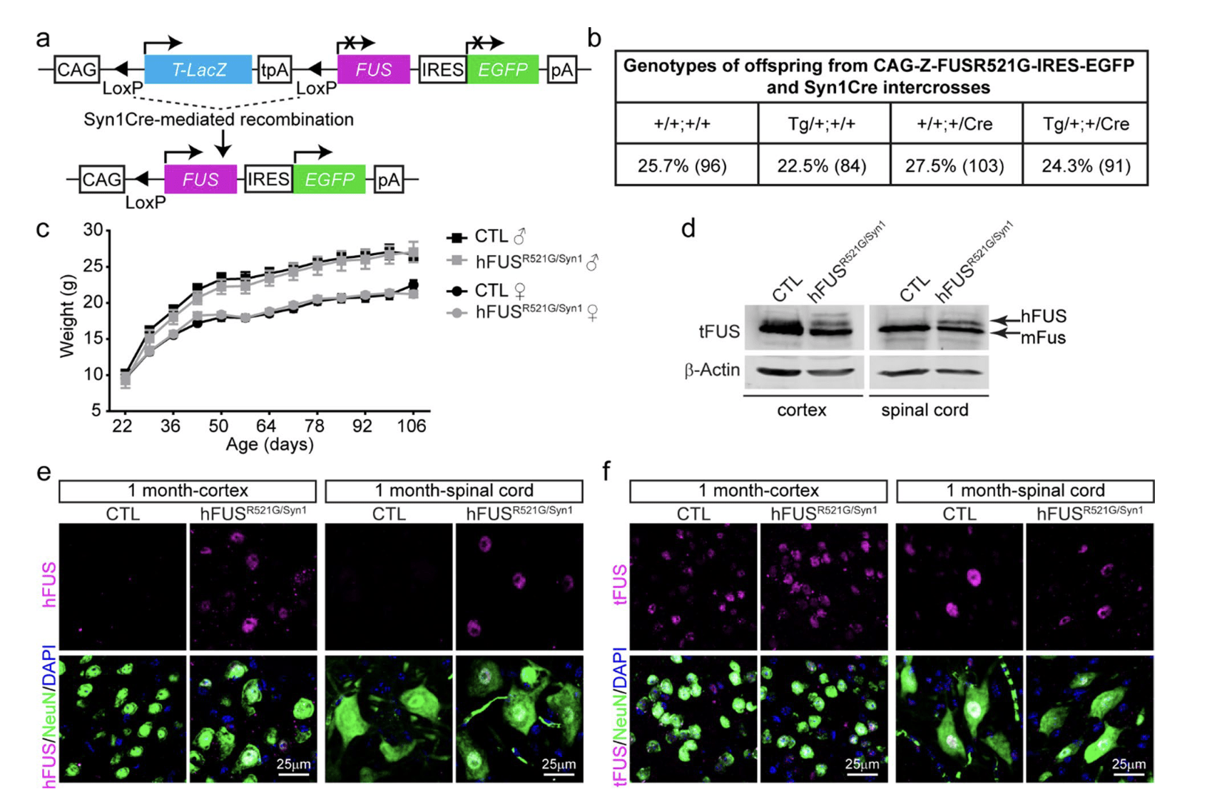
Figure 1: Generation of neuron‑specific hFUSR521G/Syn1 mice. a Schematic of human FUSR521G/Syn1Cre (hFUSR521G/Syn1) mice generation. b Mendelian ratios of offspring genotypes from 56 intercrosses between CAG‑Z‑FUSR521G‑IRES‑EGFP and Syn1Cre mice. The numbers indicated in brackets are the number of mice for each genotype. c Weight curve of hFUSR521G/Syn1 and littermate control mice 6–8 mice per group. d Immunoblot of lysates from indicated tissues of 1‑month‑old littermate control (CTL) and hFUSR521G/Syn1 mice. Proteins are detected with antibodies against total FUS (tFUS) and the loading control β‑Actin. The lower molecular weight band in the immunoblot corresponds with endogenous mouse FUS (mFus). The higher molecular weight band in the immunoblot corresponds with human FUSR521G protein (hFUS) expressed in the brain and spinal cord tissues of hFUS R521G/Syn1 mice. The brain and spinal cord of CTL and hFUSR521G/Syn1 mice stained with anti‑NeuN (neuron marker), DAPI (nuclear marker) and e anti‑hFUS or f anti‑tFUS. hFUS is only detected in NeuN positive cells in the cortex and spinal cord of FUS transgenic mice.
The study suggests that these morphological changes signal microglia and astrocytes, the immune cells of the central nervous system. While their initial arrival is protective, prolonged presence becomes toxic to neurons, reducing synaptic connections between motor neurons in the brain and spinal cord, ultimately leading to muscle atrophy and loss of motor function.
In experiments, a semi-synthetic drug derived from Withaferin A, an extract of the Ashwagandha plant, was tested. This drug effectively inhibits inflammation, allowing motor neurons to revert to a more normal state. The researchers observed neuronal regeneration in the absence of activated immune cells. This finding suggests a potential therapeutic approach for ALS by targeting inflammation to alleviate symptoms and promote neuron recovery.
Journal article: Mari Carmen Pelaez, M.C., et al., 2023. Neuronal dysfunction caused by FUSR521G promotes ALS-associated phenotypes that are attenuated by NF-κB inhibition. Acta Neuropathologica Communications.
Summary by Stefan Botha


