


The fetal immune system is not simply an immature version of the adult immune system. Even before birth, immune cells develop within distinct tissue environments, balancing immune tolerance with the capacity to respond to microbial and environmental challenges after delivery.
A new study provides a multi-tissue single-cell atlas of the late-gestation rhesus macaque fetal immune system, revealing how immune populations differ across the lung, spleen and umbilical cord blood (Figure 1).
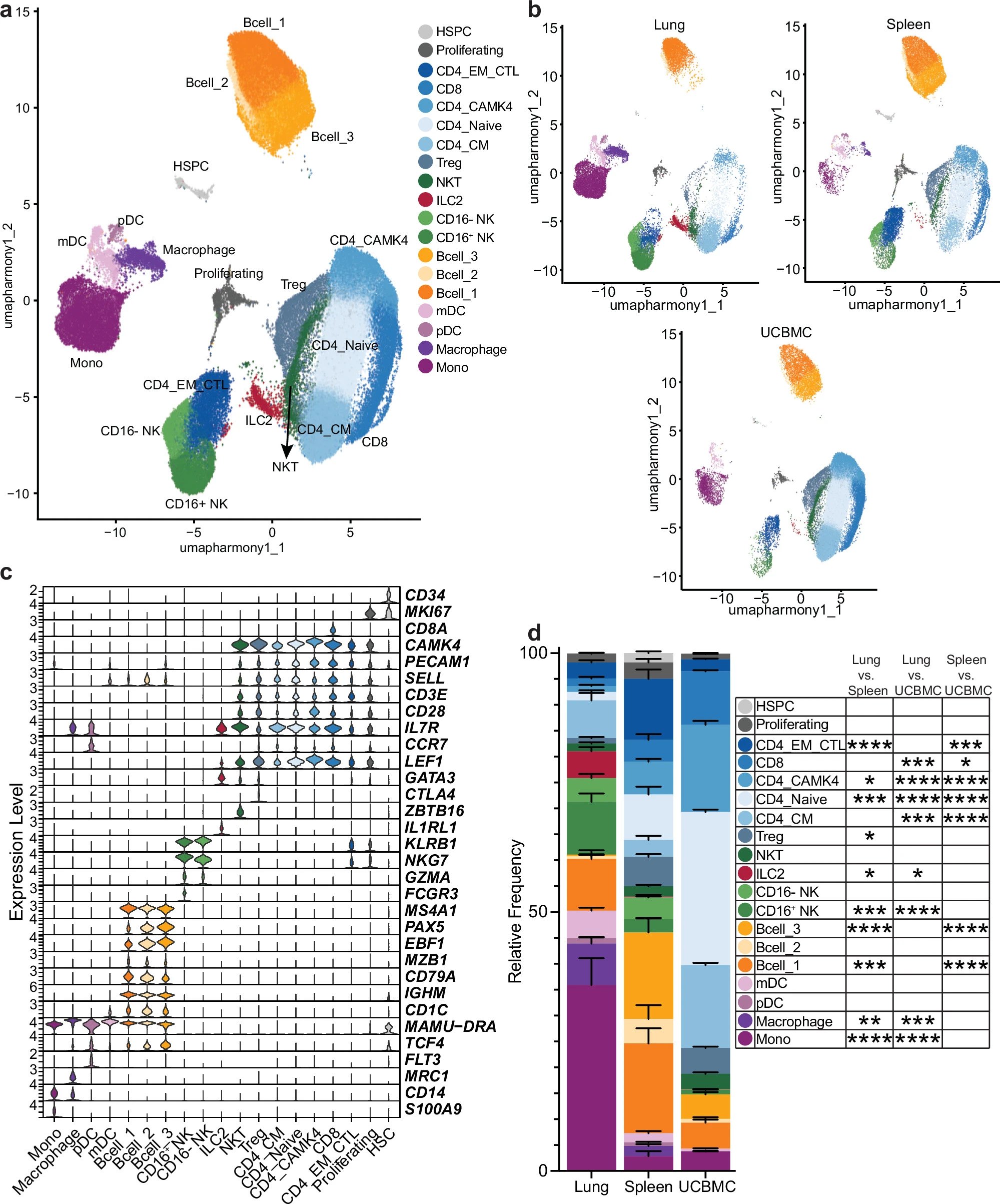
Figure 1: Single-cell analysis of fetal immune cells. a UMAP projection of 102,245 cells from the fetal lung (29,760 cells from n = 10 biologically independent animals; n = 4 female), spleen (26,504 cells from n = 6 biologically independent animals; n = 3 female), and UCBMC (45,981 cells from n = 10 biologically independent animals; n = 4 female) leukocytes of ten fetal rhesus macaques. b UMAP separated by tissue type. c Violin plots of marker gene expression used for cluster identification. d Stacked bar plots of cluster frequencies within the lung (n = 10 biologically independent animals identified via positive HTO, n = 2 HTO doublets, n = 2 HTO negative), spleen (n = 6 biologically independent animals identified via positive HTO, n = 2 HTO doublets, n = 2 HTO negative), and UCBMC (n = 6 randomly assigned pseudo-replicates from n = 10 biologically independent animals). Statistical tests were performed using a two-way ANOVA with Tukey correction in Prism (v10). Error bars represent standard error of the mean. ✳=p < 0.05, ✳✳=p < 0.01, ✳✳✳=p < 0.001, ✳✳✳✳=p < 0.0001. Cluster frequencies were calculated using n = 10 (lung) and n = 6 (spleen) positively identified HTO samples plus doublet and negative HTO frequencies from each of two pooled libraries, all treated as replicates; for UCBMC, which did not demultiplex, n = 3 pseudo-replicates were randomly assigned within each library.
Using single-cell transcriptional profiling, supported by spectral flow cytometry and functional assays, the researchers identified distinct immune landscapes in each tissue:
Fetal lung: Enriched in myeloid cells and group 2 innate lymphoid cells (ILC2s), with a more pro-inflammatory profile. Functional assays also showed higher baseline secretion of inflammatory mediators.
Fetal spleen: Dominated by T and B cells. B cells showed enhanced signatures associated with V(D)J recombination and antibody isotype switching, while CD4+ T cells displayed increased activation and a higher frequency of regulatory T cells (Tregs).
Umbilical cord blood: Predominantly composed of T cells with a more regulatory transcriptional profile. Unlike the lung, spleen and cord blood showed greater inducible responses following immune stimulation.
These findings highlight that fetal immune development is highly tissue-specific. Different compartments appear to have distinct functional capacities before birth, with some maintaining a more regulatory environment while others are poised for inflammatory responses.
By mapping immune cell populations, transcriptional programmes and intercellular communication across multiple fetal tissues, this study provides a valuable resource for understanding how immune readiness develops before birth.
Although these findings come from a non-human primate model, they offer important insight into the complexity of prenatal immune development and the foundations of postnatal immune responses.
Journal article: Doratt, B.M., et al. 2026. Single-cell atlas of fetal immunity across lung, spleen, and umbilical cord blood in nonhuman primates. Nature Communications.
Summary by Stefan Botha