


Herpes simplex virus type 1 (HSV-1), best known for causing cold sores, can alter how the brain’s resident immune cells function. New research reveals how HSV-1 infection reshapes microglial activity, potentially linking antiviral defence with impaired brain maintenance (Figure 1).
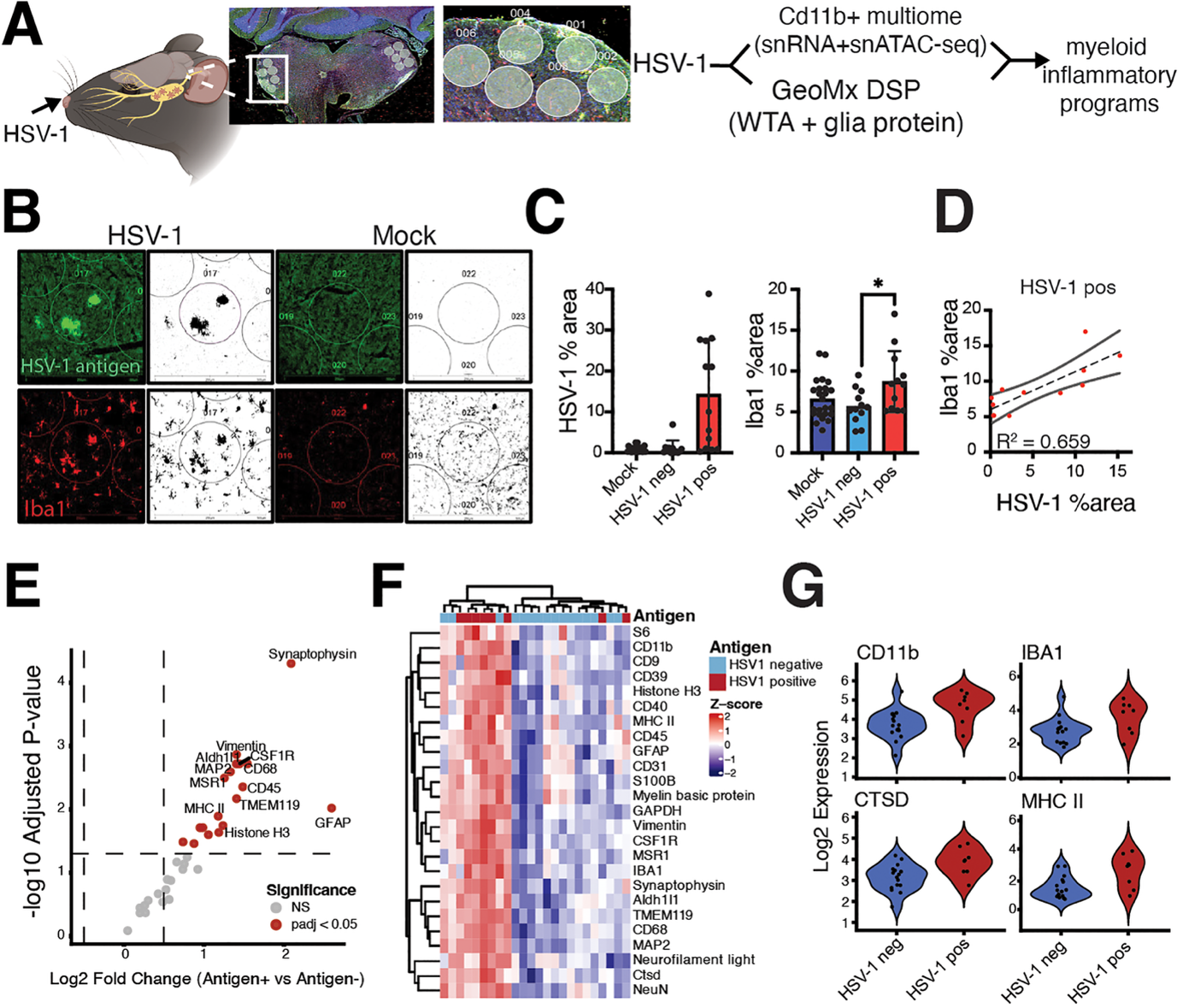
Figure 1: Spatially restricted HSV-1 infection and microglial response in the trigeminal nucleus of the brainstem. A) Adult C57BL/6J mice were intranasally infected with non-lethal HSV-1 (McKrae strain) or mock-treated and analyzed at 6 days post-infection. CD11b⁺ nuclei (n = 3 pooled populations) were isolated from whole brains for paired single-nucleus RNA-seq (snRNA-seq) and single-nucleus ATAC-seq (snATAC-seq). In a separate cohort subjected to the same infection protocol, brain sections were processed using the GeoMx Digital Spatial Profiler (DSP) with immunofluorescent labeling for HSV-1 antigen (gB) and Iba1 to guide region-of-interest (ROI) selection (24 ROIs across n = 1 animal/condition; 48 total ROIs). NanoString GeoMx Digital Spatial Profiler (DSP) Whole Transcriptome Atlas (WTA) assay were performed. Representative whole-section image from HSV-1 infection and ROI masks are shown. B) Immunofluorescence images from HSV-1-infected and mock-treated brains. HSV-1 antigen (green) localizes predominantly to the ipsilateral spinal trigeminal nucleus (SpVN) in infected animals and is absent in mock controls. Iba1 (red) signal is increased in antigen-positive regions. Grayscale images show segmentation masks used for ROI selection. C) Quantification of HSV-1 antigen-positive area (left) and Iba1-positive area (right) across mock, HSV-1 antigen-negative, and HSV-1 antigen-positive regions. Bars represent mean s.e.m.; points represent individual regions. D) Correlation between HSV-1 antigen area and Iba1-positive area in HSV-1-positive regions. Each point represents a single region; line indicates linear regression with 95% confidence interval. E) Differential protein enrichment analysis across the DSP glial protein panel comparing HSV-1 antigen-positive (HSV-1+) and antigen-negative (HSV-1-) ROIs. Protein abundance values were normalized and globally centered across ROIs prior to linear modeling. Proteins with adjusted P value < 0.05 (Benjamini-Hochberg FDR) are highlighted in red; non-significant proteins are shown in gray. (F) Per-protein Z-score–scaled normalized DSP protein abundance values across HSV-1 antigen–positive and antigen–negative ROIs. (G) Log2-normalized DSP protein abundance values for selected markers (CD11b, IBA1, CTSD, MHC II) across HSV-1 antigen-negative and antigen-positive ROIs. CD11b p = 0.0159; IBA1 p = 0.0926; CTSD p = 0.023; MHC II p = 0.00875. Fig 1A was created in BioRender.com.
Using molecular approaches to examine individual brain cells, researchers found that intranasal HSV-1 infection induced distinct microglial populations with strong type I interferon-associated antiviral responses.
At the same time, genes involved in the normal protective and maintenance functions of microglia became less active. These changes were concentrated in brain regions where the virus was detected, suggesting that HSV-1 drives localised immune remodelling.
The researchers also identified changes in chromatin, the structure that packages DNA and helps regulate gene expression. This raises the possibility that HSV-1 affects not only the immediate immune response but also the molecular programmes that determine how microglia function.
An important unanswered question is whether these changes persist after the infection has resolved or influence responses to subsequent viral reactivation.
The microglial changes observed following HSV-1 infection resemble some immune-response patterns associated with Alzheimer’s disease and other neurodegenerative processes.
Microglia are essential for defending the brain, clearing cellular debris and maintaining tissue homeostasis. If their antiviral activation compromises these functions, it could potentially contribute to an environment that makes the brain more vulnerable to damage.
This study does not establish that HSV-1 causes Alzheimer’s disease. Instead, it identifies a potential mechanism connecting viral infection with altered microglial function. Whether these changes persist and contribute to neurodegeneration over time remains to be investigated.
Understanding how infections reshape the brain’s immune environment could help explain why some individuals are more susceptible to neurodegeneration, particularly in the context of ageing and other risk factors.
Journal article: Frietze S, et al. 2026. Intranasal HSV-1 infection drives region-specific interferon-dominant microglial remodeling. PLOS Pathogens.
Summary by Stefan Botha