


Researchers have uncovered previously unrecognised diversity in the way resident macrophages develop in the peritoneal cavity, identifying two distinct pathways that generate the large cavity macrophages (LCMs) that help maintain tissue health and defend against infection (Figure 1).
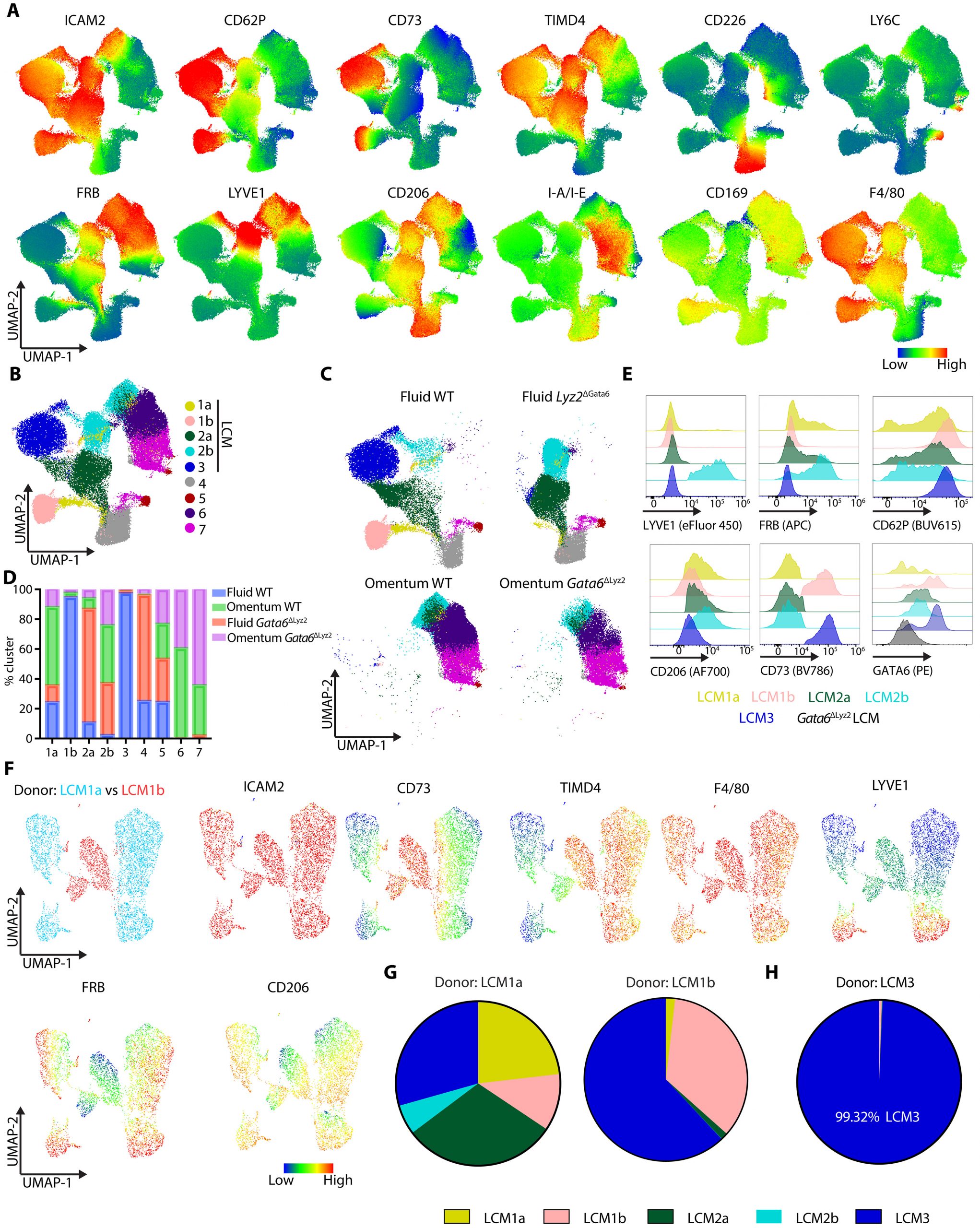
Figure 1: Large peritoneal macrophages comprise distinct subsets along two differentiation trajectories. (A) UMAP of pregated peritoneal fluid macrophages (CD11b+CD115+) and omental macrophages (CD64+F4/80+) from multicolor spectral flow cytometry, overlaid with normalized expression of the indicated markers. Color scale indicates relative expression level (low to high). (B) Unsupervised clustering of the same UMAP identifies nine macrophage clusters. (C) UMAPs split by group showing the distribution of peritoneal fluid WT, fluid Gata6ΔLyz2, omentum WT, and omentum Gata6ΔLyz2 cells. (D) Percentage contribution of each sample group to clusters 1a to 7. Colors indicate fluid WT (blue), fluid Gata6ΔLyz2 (salmon), omentum WT (green), and omentum Gata6ΔLyz2 (purple). (E) Representative contour plots comparing key markers (ICAM2, LYVE1, FRβ, CD206, CD62P, CD73, and GATA6) across the identified LCM subsets. Data in (A) to (E) were generated from 8- to 11-week-old male mice, including n = 7 WT and n = 4 Gata6ΔLyz2 mice. (F) Competitive adoptive transfer of sorted LCM1a and LCM1b cells (1:1), analyzed on day 8 by spectral flow cytometry and unsupervised analysis of pre-gated CD11b+CD115+ peritoneal fluid macrophages. Left, UMAP overlay of LCM1a-derived versus LCM1b-derived cells. Right, marker-expression overlays (ICAM2, CD73, TIMD4, F4/80, LYVE1, FRβ, and CD206) defining donor cell identity. Color scale indicates relative expression level (low to high). (G) Subset distribution among donor-derived cells at day 8, by LCM1a (left) and LCM1b (right) donor: LCM1a (orange), LCM1b (red), LCM2a (green), LCM2b (cyan), and LCM3 (blue) cells. Individual data points and statistical analysis are shown in fig. S1C. Adoptive transfer experiments in (F) and (G) used donor cells isolated from 11- to 15-week-old female ActinmTFP and Ms4a3Cre x TdTomato mice and transferred into n = 3 7- to 24-week-old female recipient mice. (H) Adoptive transfer of sorted LCM3 cells isolated from 11-week-old male Ms4a3Cre x TdTomato mice followed by day 8 analysis in n = 3 8-week-old male recipient mice.
The findings challenge the traditional view that resident macrophages arise through a simple division between embryonic and adult-derived cells. Instead, the study shows that even macrophages with similar identities can develop through different molecular routes and adopt distinct functions.
The peritoneal cavity contains several specialised macrophage populations. Among the most abundant are large cavity macrophages, or LCMs, which reside in the fluid surrounding the abdominal organs and contribute to immune surveillance and tissue homeostasis.
Using fate-mapping experiments in mice, researchers identified two separate pathways capable of producing these cells.
The dominant pathway generates Gata6-positive LCMs through a Gata6-dependent differentiation programme. This pathway remained functional even when the researchers experimentally increased macrophage turnover, suggesting that it represents a robust mechanism for maintaining the resident population.
A second, quantitatively smaller pathway was found to originate from monocytes. These cells developed into a population of LYVE1-positive LCMs, but produced relatively few Gata6-positive cells.
Unlike the dominant pathway, the alternative route did not require Gata6. Instead, it depended on a regulatory region within the Zeb2 gene, a transcriptional regulator involved in monocyte development.
When researchers used mice carrying mutations that disrupt this Zeb2 enhancer, the monocyte-derived pathway was severely impaired. The Gata6-dependent pathway, however, remained largely unaffected.
This demonstrates that apparently similar resident macrophages can be generated through fundamentally different developmental programmes.
The researchers also found that the smaller monocyte-derived pathway has a distinct functional role. These LCMs were particularly effective at surveying the mesothelial surface, the thin layer of cells lining the abdominal cavity. When an available niche appeared among the macrophages associated with this surface, the monocyte-derived LCMs could move in and replenish the population of mesothelial border macrophages.
This behaviour was not simply a consequence of their location. It suggests that the developmental pathway through which macrophages arise can influence where they go and what functions they perform.
Macrophages have traditionally been classified according to whether they originate from embryonic precursors or adult haematopoietic cells. The new findings suggest that this distinction does not capture the full complexity of macrophage development.
Instead, different precursor populations can generate macrophages with overlapping characteristics through distinct transcriptional programmes, ultimately producing cells with different tissue-surveillance roles.
Understanding these developmental pathways could be important for studying diseases in which macrophage populations contribute to inflammation, tissue repair or immune regulation.
The findings highlight that macrophage identity is shaped not only by where a cell comes from, but also by the molecular pathway it follows to become a resident immune cell.
Journal article: Han, J, et al. 2026. Two differentiation pathways generate large peritoneal macrophages with one replenishing mesothelial border macrophages. Science Immunology.
Summary by Stefan Botha