


HLA-DR is a molecule typically expressed by antigen-presenting cells and is associated with antigen presentation. Interestingly HLA-DR is also expressed on T cells and has been associated with T cell activation. However, why and how activated T cells express HLA-DR is not fully understood. A recent study by Tippalagama et al. aimed to characterize the phenotype of effector CD4 T cells in active Tuberculosis.
Transcriptomic profiling of non-naïve CD4 T cells from M.tb uninfected, M.tb infected and individuals with active TB identified significantly higher expression levels of molecules associate with antigen-specific T cell activation and inflammation, and specifically identified higher expression of HLA-DRB1 in TB patients compared with both M.tb uninfected and infected individuals. As observed by others, researchers demonstrated higher frequencies of HLA-DR+ circulating M.tb-specific CD4 T cells in tuberculosis patients compared with both M.tb uninfected and infected individuals. Further, HLA-DR+ M.tb-specific CD4 T cells predominantly expressed phenotypic markers associated with an effector memory phenotype (CD45RA-CCR7-) and increased expression of cytotoxic molecules and proinflammatory cytokines (TNF). Lastly, they demonstrated that proliferating T cells express high levels of HLA-DR, suggesting that HLA-DR could be used as a proxy to identify recently divided CD4 T cells upon M. tuberculosis Ag exposure.
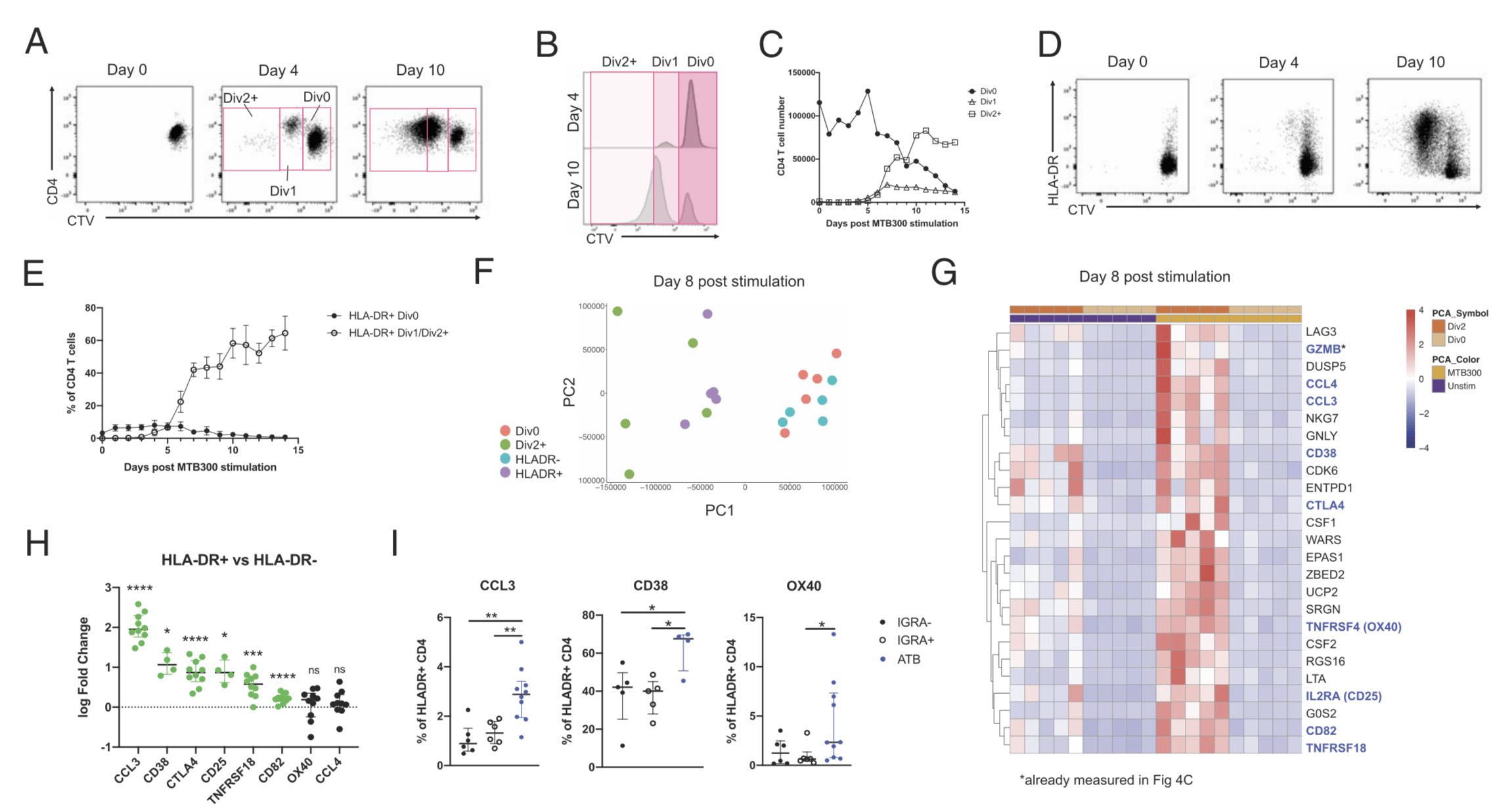
HLA-DR expression marks recently divided CD4 T cells upon M. tuberculosis Ag exposure. Using our in vitro model of M. tuberculosisspecific T cell proliferation assay, PBMC of healthy IGRA1 individuals were stained with proliferation dye CTV and in vitro stimulated with the MTB300 peptide pool (28) for 14 d. Each day post stimulation, CD4 T cells were identified as either undivided (Div0), divided once (Div1), or divided more than once (Div2+) by flow cytometry. (A) Representative co-staining plots of CD4 and CTV expression before (day 0), 4 d, and 10 d post-MTB300 stimulation within CD4 T cells. (B) Representative histogram of CTV expression at 4 d and 10 d post-MTB300 stimulation. (C) Number of CD4 T cells within each CTV division category in function of days post MTB300 stimulation. (D) Representative co-staining plots of HLA-DR and CTV expression before (day 0), 4 d, and 10 d postMTB300 stimulation within CD4 T cells. (E) Frequency of HLA-DR1 undivided (Div0) and HLA-DR+ divided (Div1/Div2+) CD4 T cells in function of days post-MTB300 stimulation. (A-E) Combined data from n = 5 IGRA+ individuals. (F) Principal component analysis based on all genes expressed in sorted CD4 T cell populations and (G) heatmap representing the expression of the top 25 genes upregulated in Div2+ versus Div0 non-naive CD4 T cells upon MTB300 stimulation. Genes highlighted in blue have a flow cytometric Ab available for measurement of their protein expression. Cells were sorted from 5 IGRA1 individuals at day 8 poststimulation with either DMSO or MTB300 (see gating strategy in Supplemental Fig. 1B), and their transcriptomic profile determined by RNA-seq. (H) Fold change in positive frequency of selected markers to identify recently divided cells [blue genes in (G), excluding GZMB] in HLA-DR1 compared with HLA-DR- CD4 T cells in ATB patients (n = 10 for CCL3, CTLA4, TNFRSF18, CD82, OX40, and CCL4; n = 4 for CD38 and CD25) determined by flow cytometry. Significant markers (p < 0.05) are represented in green. (I) Frequency of positive cells for CCL3, CD38, and OX40 in HLA-DR1 CD4 T cells of ATB [similar cohort to (H)], IGRA1 (n = 6) and IGRA (n = 6) individuals. *p < 0.05, **p < 0.01, *** p < 0.001, **** p < 0.0001, nonparametric paired Wilcoxon test. (Source: Tippalagama et al. 2021)
Researchers concluded HLA-DR expression is a marker of such effector cells and that HLA-DR+ CD4 T cells were increased in individuals with ATB compared with M.tb uninfected and infected individuals. Further, they suggest that HLA-DR might be a useful marker for identifying effector T cells and monitoring immune responses not only in the context of TB but also in many other infection and vaccination models.
Journal Article: Tippalagama et al. 2021. HLA-DR Marks Recently Divided Antigen-Specific Effector CD4 T Cells in Active Tuberculosis Patients. Journal of Immunology
Summary by Cheleka AM Mpande


