


Lung functional decline is one of the outcome measures looked at when it comes to patients with chronic obstructive pulmonary disease (COPD). What could go a long way in identification, better classification, management and monitoring of COPD patients are biomarkers that can help determine changes in forced expiratory volume (FEV1) and thus provide support on the best possible therapeutic approaches to put in place for each COPD phenotype, based on the level to which lung function declines.
The avenue that Di Stefano and colleagues took was aimed at determining expression markers of inflammation within different categories of patients with COPD, that is, those with rapid, slow or no lung functional decline. Functional decline in these patients was measured and followed up for a period of 5.8 years.
Results from immunohistochemistry done on the bronchial epithelium (figure 1) showed that the numbers of several inflammatory cells flooding into the bronchial epithelium were similar across all three groups of COPD patients looked at within this study. Total Immunoglobulin A (IgA) and polymeric Ig receptor (pIgR) did not differ much either however, when it came to the secretory IgA score, this was seen to reduce in rapid decliners in comparison to non-decliners.
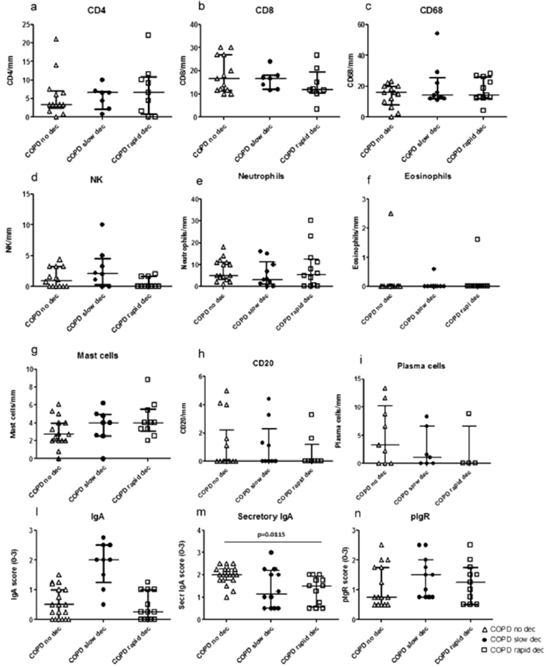
Figure 1: “Quantitation of CD4 (a), CD8 (b), CD68 (c), natural killer cells (d), neutrophils (e), eosinophils (f), mast cells (g), B cells (CD20) (h), plasma cells (i), total IgA (l), secretory IgA (m) and polymeric immunoglobulin receptor (pIgR) (n) in the bronchial epithelium of non-decliners (n = 21), slow decliners (n = 14) and rapid decliners (n = 15) with COPD. Bar represents median value. Secretory IgA was significantly reduced in rapid decliners compared to non-decliners. Exact p value is reported in the graph (m)”
The authors also noted that there was a decreased plasma cell count in the lamina propria of rapid decliners compared to non-decliners (figure 2). Di Stefano et al believe that the data would point to an impaired, or somewhat altered humoral response being a contributor to the rapid decline in lung function that ends up exacerbating COPD in patients that already have it. The results reported on in this paper are also said to be in line with other literature that suggests a link and correlation between a drop in IgA or pIgR and COPD.
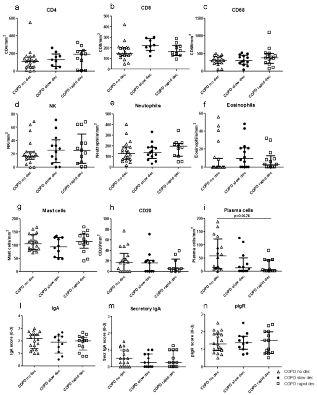
Figure 2: “Quantitation of CD4 (a), CD8 (b), CD68 (c), natural killer cells (d), neutrophils (e), eosinophils (f), mast cells (g), B cells (CD20) (h), plasma cells (i), total IgA (l), secretory IgA (m) and polymeric immunoglobulin receptor (pIgR) (n) in the bronchial lamina propria of non-decliners (n = 21), slow decliners (n = 14) and rapid decliners (n = 15) with COPD. Plasma cell numbers were significantly reduced in rapid decliners compared to non-decliners. Exact p value is reported in the graph (i). Bar represents median value”
Based on the aforementioned information, the authors had this to say; “independently of the degree of severity of bronchial obstruction, the lower levels of secretory IgA in epithelium and plasma cells in lamina propria that we found in rapid decliners vs. non-decliners are a specific feature of functional decline and may be considered as markers of lung function decline in COPD patients.”
What would need to be studied further now are the mechanisms governing the development of plasma cells in order to uncover how these drive the reduction of plasma cells and secretory IgA in rapid decliners with COPD.
Journal article: Di Stefano, A., et al., 2022. Decreased humoral immune response in the bronchi of rapid decliners with chronic obstructive pulmonary disease. Respiratory Research.
Summary by Vanessa Muwanga


