


In a recent paper, researchers have described a promising approach to identify individuals at risk of developing type 1 diabetes, a severe autoimmune disease, by analyzing T cells in the blood (Figure 1). The scientists focused on T cells, a key player in autoimmune responses, and isolated them from both human and mouse blood samples. By closely studying the T cells associated with type 1 diabetes, they successfully differentiated individuals with active autoimmunity, indicative of diabetes risk, from those without significant autoimmunity. Remarkably, this differentiation was achieved with 100% accuracy in a small sample group.
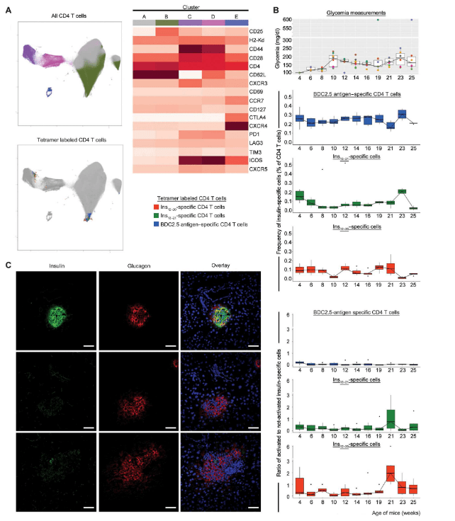
Figure 1: Following the state of activation of circulating antigen-specific CD4 T cells in NOD/LtJ mice by 22-color multiparametric flow cytometry. In this representative experiment, eight mice were followed from week 4 to 25 and sampled for blood every other week. (A) All CD4 T cells from all mice at all time points were analyzed and split in groups by unsupervised K-means clustering. Visualization of clusters is shown in graphical form by UMAP (top) and heatmap representation (right). The positioning of pMHC tetramer+ cells (orange for Ins12–20-, green for Ins13–21-, and blue for BDC2.5 antigen–specific T cells) in the general UMAP (gray color) is shown in the bottom. (B) Plots of frequency and activation state for each tetramer+ population, BDC2.5 antigen–specific, Ins12–20, and Ins13–21. Activation state was defined by the ratio of cells retained in clusters C and D versus cells in A and B. Glycemia is shown in the top; it shows the synchrony between hyperglycemia and the activation of the Ins12–20 population. (C) Nonclinically diabetic mice at week 25 have had islet autoimmunity. Representative immunofluorescence images of the pancreas of the three mice that were not diabetic at the end of study. Most of the islets were either intact and producing both insulin and glucagon or totally devoid of insulin-producing cells. Rare islets were still showing signs of insulitis (bottom). A precise quantitation of the three phenotypes could not be performed because of the small number of animals. Scale bars, 50 μm.
This discovery marks a significant advancement, as it offers the prospect of identifying the autoimmune process responsible for type 1 diabetes at an early stage, providing an opportunity to intervene and prevent or significantly delay the onset of the disease.
The study’s innovative methodology involved constructing protein complexes mimicking the immune proteins and insulin fragments recognized by specialized T cells called CD4 T cells to initiate the autoimmune reaction. By capturing anti-insulin CD4 T cells from blood samples and analyzing their gene activity and protein expression, the researchers created a classification algorithm that accurately identified individuals at risk of ongoing anti-islet autoimmunity.
If further research successfully refines this method for identifying at-risk individuals and monitoring their autoimmunity status, it could not only facilitate timely treatment but also allow for disease progression monitoring and evaluation of new preventive therapies. This breakthrough holds immense potential for transforming the landscape of type 1 diabetes management and prevention. This innovative method, if validated through further research, has the potential to select suitable candidates for treatment that could halt the autoimmune process responsible for type 1 diabetes, effectively making it a preventable condition.
Journal article: Sharma, S., et al. 2023. Measuring anti-islet autoimmunity in mouse and human by profiling peripheral blood antigen-specific CD4 T cells. Science Translational Medicine.
Summary by Stefan Botha


