


Patients with systemic lupus erythematosus (SLE) and coronavirus disease 2019 (COVID-19) share common features such as a dysregulated type I interferon response, increased risk of thromboembolism, and activation of the classical complement pathway. Many of these symptoms are thought to be mediated by an imbalance in neutrophil extracellular trap (NET) formation and degradation.
Some studies have already been published on the impact of NET formation in patients with COVID-19 and SARS-CoV-2 infection on autoimmune diseases such as SLE. Knopf and colleagues now assessed the serological changes in the serum of patients with SLE with regards to NET formation in samples taken before and during the pandemic (Figure 1). They investigated the presence of cell-free DNA, Myeloperoxidase (MPO)-DNA and Neutrophil Elastase (NE)-DNA complexes, and the activity of NE in serum of 100 SLE patients from a recently described Swedish cohort.
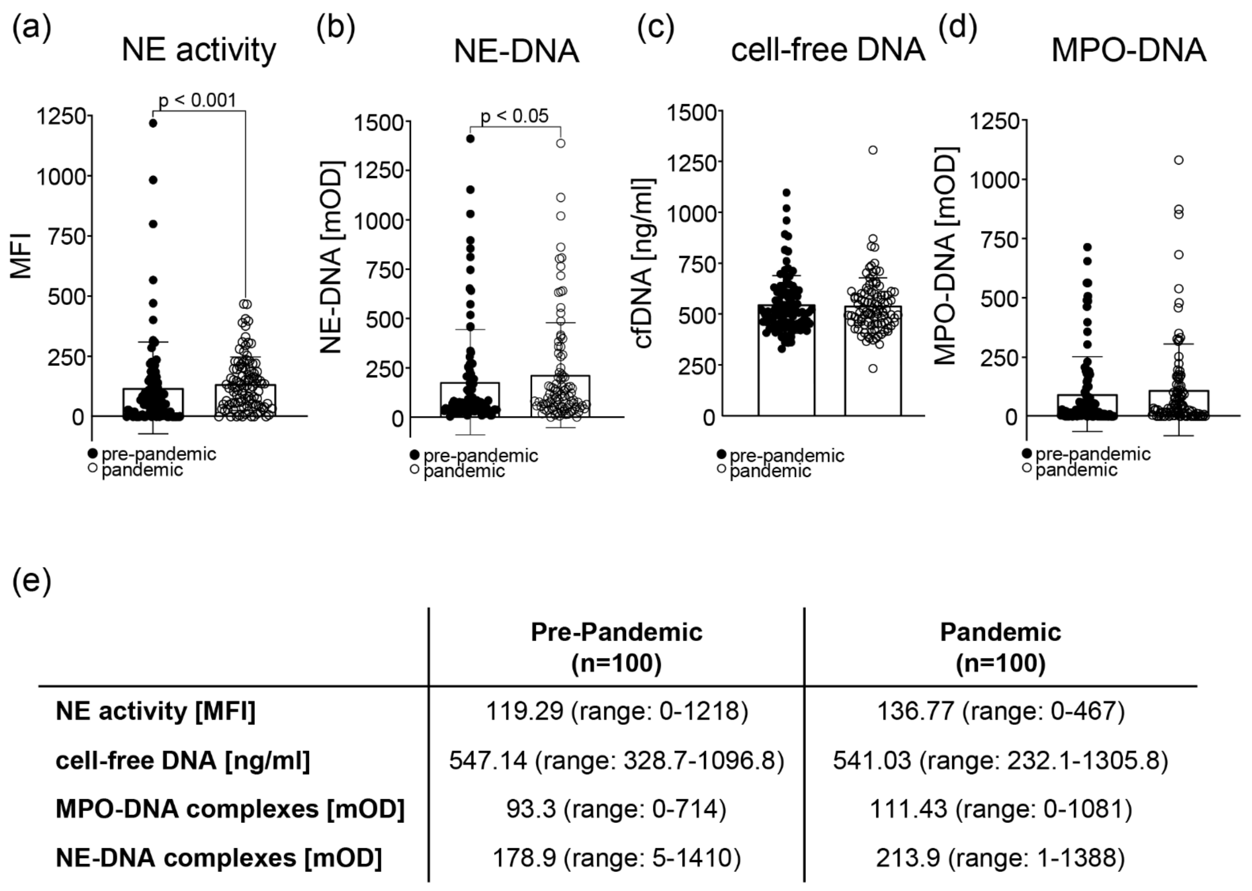
Figure 1: Markers for neutrophil extracellular traps (NETs) in the sera of patients with SLE pre-pandemic compared to the pandemic. (a) The activity of neutrophil elastase (NE) was assessed by the increase in the mean fluorescence intensity (MFI) after the conversion of a specific fluorescent substrate. The activity of the NE was significantly higher (p < 0.001) in the pandemic sera. (b) The presence of NE-DNA complexes was significantly different (p < 0.05) between the pre-pandemic and pandemic samples, as analyzed by ELISA. (c) The presence of cell-free DNA in the serum of patients was measured by a Quant-iT PicoGreen dsDNA Assay Kit, and no significant differences were observed between the different time points. (d) The presence of MPO-DNA complexes was analyzed by ELISA, showing no significant differences between the pre-pandemic and the pandemic cohorts. (e) Mean values and ranges for all three NET parameters were analyzed (NE activity, cell-free DNA, and MPO-DNA complexes) in the sera of SLE patients: pre-pandemic vs. pandemic. Statistical significance was calculated using the Wilcoxon matched-pairs signed rank test.
The activity of NE and the presence of NE-DNA complexes was significantly higher in the serum from the pandemic period compared to pre-pandemic samples. The NE activity also correlated with other clinical parameters such as hemoglobin concentration, neutrophil count, and levels of complement protein 3 and 4. When the pandemic samples were further divided according to their serological positivity for exposure to SARS-CoV-2, the same correlations were found and the levels of cell-free DNA were inversely associated with malar rash (ACR 1). Unfortunately, there was no correlation with other clinical markers. It would have been advantageous to also compare the presence and activity of NET markers in the serum of healthy donors but it was impossible to obtain enough longitudinal serum samples from healthy donors during this time. Nevertheless, this study is the first to our knowledge to report changes in NET formation in the same person over time.
Journal article: Knopf, J, et al., 2022. NET Formation in Systemic Lupus Erythematosus: Changes during the COVID-19 Pandemic. Cells.
Summary by Jasmin Knopf


