


In a recent study, researchers investigate immune cell behavior during human lung development in embryos and fetuses (Figure 1). While previous research has extensively covered immune cell functions in various tissues, the understanding of their processes in lung development has been limited.
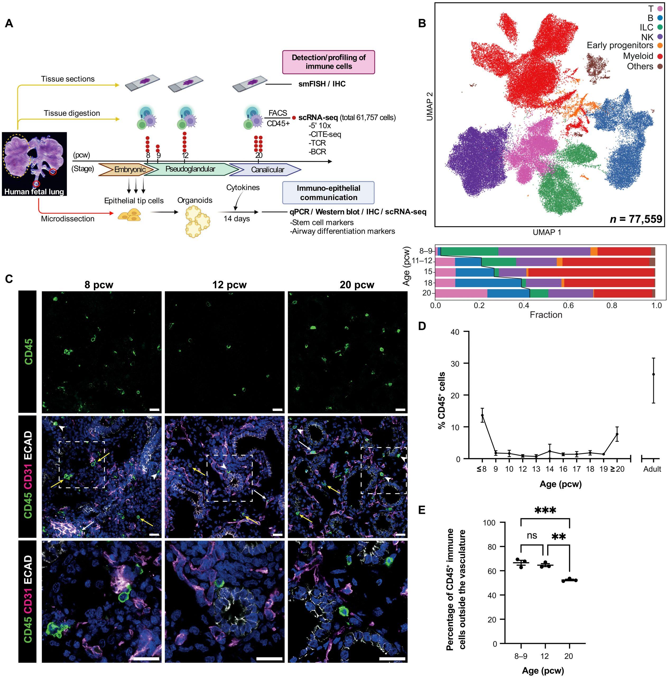
Figure 1: Immune cells are abundant in human fetal lungs. (A) Experimental overview. Human fetal lung tissue was digested and FACS-sorted to isolate CD45+ immune cells for scRNA-seq (red dot, biological replicate; n = 19; FACS gating strategy: fig. S1B). Tissue sections across developmental stages were used for cell type validation (IHC and smFISH), whereas embryonic tissue was used to generate organoids for functional studies. (B) UMAP (top) and bar chart (bottom) colored by broad cell populations in the single-cell datasets. Representative IHC images (C) show the spatial distribution of CD45+ immune cells within the endothelium (CD31+, white arrows), epithelium (ECAD+, arrowheads), and mesenchyme (yellow arrows) during fetal lung development (blue, DAPI+ nuclei; scale bar, 20 μM). The proportion of immune cells, as a percentage of all DAPI+ nuclei, was quantified in cryosections at weekly time points throughout lung development (D) using ImageJ. Data are presented as means ± SEM, n ≥ 3 biological replicates. (E) The proportion of CD45+ immune cells outside the CD31+ vasculature versus inside was calculated at 8 to 9, 12, and 20 pcw (mean ± SEM, n = 3 biological replicates). **P < 0.01 and ***P < 0.001 were calculated by one-way ANOVA followed by Tukey’s post hoc test.
The objective of this study was to track the progression of the fetal immune system and its potential impact on embryonic lung development. Utilizing advanced techniques like single-cell RNA sequencing (scRNA-seq), immunohistochemistry, and additional assays, the team observed notable variations in immune cell populations across different fetal developmental stages.
The early stages were marked by the prevalence of progenitor and innate immune cell populations. However, as development advanced, these cell types were replaced by T- and B-lymphocytes. CD45+ cells, were consistently present across all developmental stages and lung-associated tissue regions, displaying varying quantities based on time and location.
A key finding was the identification of B-cell maturation within embryonic lungs. The study also revealed widespread production of Interleukin-1 beta (IL-1β) by myeloid cells, a factor influencing epithelial stem cell differentiation.
By combining transcriptomic analyses with immunohistochemistry, the study provided insights into the structural and functional roles of immune cells during fetal and embryonic development. The interactions of immune cell-secreted cytokines, validated to impact epithelial cell differentiation, underscore the dual role of immune cells in both defence and lung epithelial development during fetal development, supporting earlier hypotheses. These findings challenge established notions, enriching our understanding of the dynamic interplay between immune cells and lung development.
Journal article: Barnes, J. L., et al. (2023). Early human lung immune cell development and its role in epithelial cell fate. Science Immunology.
Summary by Stefan Botha


