


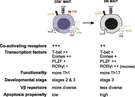
Properties and relationship of the main MAIT cell subsets. (Source: Dias et al., 2018 PNAS)
Mucosal associated invariant T (MAIT) cells are MHC-class I-related restricted cells found in low frequencies in blood. Studies have shown a decrease in MAIT cell blood frequencies during infection. It is unclear whether depletion of MAIT cells in the blood is due to apoptosis or migration to sites of infection, and whether MAIT cells in blood and tissue have distinct phenotypic and functional characteristics. Voillet et al., aimed to contribute to this knowledge gap by determining if MAIT cells from tissue have distinct characteristics from MAIT cells found in the blood.
Researchers used flow cytometry and transcriptomic analysis to compare MAIT cell phenotypes in the blood and thoracic duct lymph (TDL). Cells in the TDL are from different sites of the body, therefore MAIT cells detected in this compartment reasonably estimate tissue-specific cells. Expression of CCR6 and CXCR3 was higher in MAIT cells from the TDL compared with blood, whereas the expression of CCR4 and CCR7 were similar between the two compartments. This confirms that MAIT cells detected in the TDL are truly tissue-specific cells. Transcriptional analysis showed an enriched IFN signature in blood MAIT cells compared to TDL MAIT cells. Finally, they showed similar TCRβ clonal diversity of sorted Vα7.2+MAIT cells from blood and TDL. This suggests that at least in healthy individuals there is no tissue-specific enrichment of specific clonotypes.
Overall these results demonstrate that MAIT cells are detectable in the lymph and can exit tissue. MAIT cells in the TDL and blood have different transcriptional capacity. Further studies would be required to determine if functional capacity differ, as well as determine if MAIT cell depletion in the blood is due to migration to the site of infection.
Journal Article: Viollet et al., 2018. Human MAIT cells exit peripheral tissues and recirculate via lymph in steady state conditions. JCI Insight


