


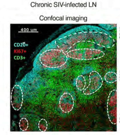
A representative confocal image of follicular cells from 1 chronically SIV-infected animal. Germinal centres (GC) were defined by CD20+Ki67+ coexpression, and CD4+ (CD3+CD4+) and CD8+ (CD3+CD4–) T cells were quantified within each GC. Scale bar: 400 μm. (Source Ferrando-Martinez et al., 2018)
CD8 T cells have been shown to be one of the main cellular mediators of HIV/SIV control. One of the major sites of HIV/SIV viral replication and persistence is lymph node. Understanding the dynamics of CD8 T cell anti-HIV immunity in the lymph node could aid in the development of future cure strategies.
Ferrando-Martinez et al., showed that SIV-specific follicular (f)CD8 T cells are observed in both intact and disorganised lymph node follicles, and accumulate during chronic infection. SIV-specific fCD8 T cells did not have superior cytokine co-expression of IFN-γ, TNF and MIP-α capacity compared with non-fCD8 T cells. However, researchers observed significantly higher expression of granzyme (Grz) A, GrzB and perforin, as well increased cytotoxic activity of SIV-specific fCD8 compared with non-fCD8 T cells detected in the lymph node.
Researchers hypothesised that increased fCD8 T cells could be due to high viral replication, as CD8 T cells have been shown to be the main mediators of viral control in. To their surprise fCDC8 T cells were not preferentially colocalised with SIV-RNA+ cells using both confocal microscopy and histocytometry. Suggestin that though SIV replication is necessary for fCD8 accumulation, it may not be sufficient to induce T cell trafficking. Using an Africa green monkey*- SIV infection models, researchers showed that activation of cells plays an important role in fCD8 T cells trafficking. Additionally, this activation was also associated with increased proportions of CXCL10 expressing monocytes. Majority of CD8 T cells observed in the lymph node expressed high levels of CXCR3, the receptor for CXCL10. This suggest a potential mechanised of CXCL10-CXCR3 mediated attraction of fCD8 T cells.
In summary, this results validated the use of non-human primate models to study the dynamics of fCD8 T cells in the lymph node during SIV infection. Additionally, they demonstrated a potential role of activation and inflammation in accumulation of cytotoxic SIV-specific fCD8 T cells. Justifying the exploration of fCD8 T cells as one of the avenues for achieving HIV cure.
*African Green Monkeys have very low activation in the lymph node
Journal Article: Ferrando-Martinez et al., 2018. Accumulation of follicular CD8+ T cells in pathogenic SIV infection. Journal of Clinical Investigation.
Article by Cheleka AM Mpande


