Improved Tetramer Staining Protocol Rius et al., 2018. Adaptation of Figure 1 (Journal of Immunology)
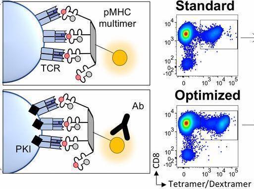
Tetramers enable detection of T cells directly ex-vivo without the need of in vitro T cell stimulation. However, tetramers staining require strong T cell receptor (TCR) and tetramer binding. This leads to the underestimation of the T cell repertoire, as functional T cells with low affinity TCR’s or recently activated T cells that have down regulated TCR expression are not bound. The development of higher order multimer peptide-major histocompatibility complex molecules such as dextramers (10 molecules) and dodecamers (12 molecules) enable detection of T cells missed by conventional tetramers. However, these molecules are not as widely available as tetramers.
Rius et al., aimed to determine an optimized tetramer staining protocol that enabled detection of T cells with low affinity TCR’s or low expression of TCRs. They optimised tetramer staining by using dasatinib (protein kinase inhibitor) to inhibit TCR downregulation and antibodies that bind to the flourochrome the tetramer was conjugated to.
Researchers showed that though the optimized protocol did not increase the overall frequency of tetramer (tet)+ CD8+ T cells, the protocol did result in increased fluorescent intensity and better separation between tet+ and tet- CD8 T cells. This was attributed to lower off-rate (dissociation) between the TCR and bound tetramers. A major caveat of tetramers is the underestimation of functional T cells. Researchers had previously shown data on individuals whom had functional T cell responses to peptide loaded to tetramers but did not bind to the tetramer using the standard tetramer staining protocol. Using the optimized tetramer staining protocol they were able to detect tet+ CD8 T cells, in these individuals. Additionally, they demonstrated that the optimised protocol resulted detection of an increased TCR diversity. Finally, Rius et al., also showed that the improved staining protocol resulted in improved detection of yellow fever virus-specific T cells, as well as detection functional tumour-specific T cell clones that were missed by standard tetramer staining protocols.
In summary, Rius et al., have shown that simply optimising current tetramer staining protocol without the use of dextramers and dodecamers results in improved detection of antigen-specific T cells with low TCR affinity. Additionally, they showed detection of increased TCR diversity compared to standard tetramer staining protocols.
Journal Article: Rius et al., 2018. Peptide−MHC Class I Tetramers Can Fail To Detect Relevant Functional T Cell Clonotypes and Underestimate Antigen-Reactive T Cell Populations. Journal of Immunology.
Article by Cheleka AM Mpande