


Researchers have discovered that microglia, the brain’s resident immune cells, play a critical role in rebuilding neural connections as the brain recovers from ketamine anaesthesia (Figure 1). The study also reveals striking differences between female and male brains, with recovery-associated neural plasticity occurring predominantly in females.
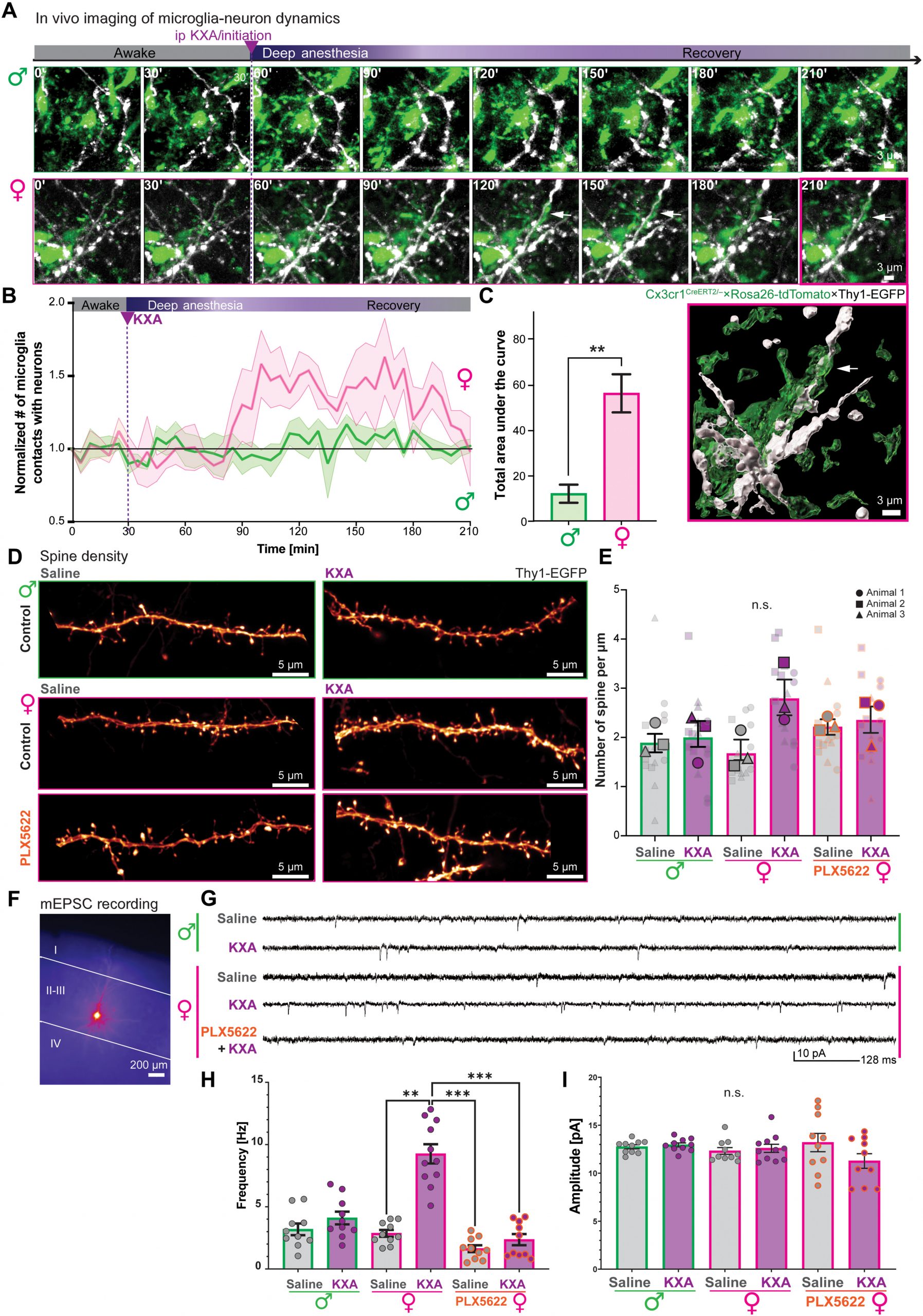
Figure 1: Female microglia interact with neurons, promoting spinogenesis and plasticity upon ketamine recovery. (A to C) In vivo two-photon imaging in the primary visual cortex (VISp) through a cranial window of Cx3cr1CreERT2/−×Ai9×Thy1-EGFP mice of both sexes spanning the phases of awake, deep anesthesia, and recovery after KXA administration. (A) Sequential snapshots of microglia (green) and excitatory neurons (white). Top, males; bottom, females; white arrow, prolonged microglia-dendrite contact; magenta frame, 3D surface rendering of the 210-min frame in females. Scale bars, 3 μm. (B) Normalized number of microglia and Thy1-EGFP neuronal process contacts over time in males (green) and females (magenta) as mean ± SEM confidence band. Dashed green line, KXA injection. Five animals per condition. (C) Bar chart of the mean total area under the curves in (B). Mean ± SEM of five animals per condition. Unpaired t test with Welch’s correction, **P < 0.01. (D and E) Spine density quantification in VISp, layer II/III in Thy1-EGFP mice 4 hours after saline or KXA injection. (D) Representative super-resolution images of dendritic processes. Top, males; middle, females; bottom, 1.5-week PLX5622-treated females to deplete microglia. Scale bars, 5 μm. (E) Bar chart of the mean number of spines per micrometer with ±SEM. Five dendrites per animal. Symbols, different animals. Three animals per condition. One-way nested analysis of variance (ANOVA), not significant (n.s.) P > 0.05. (F to I) mEPSC (miniature excitatory postsynaptic currents) recording from layer II/III VISp pyramidal neurons, 4 hours after saline or KXA injection in males, females, or microglia-depleted females (PLX5622). (F) Representative epifluorescence image of a biocytin-filled pyramidal neuron after mEPSC recording. Scale bar, 200 μm. (G) Representative recorded mEPSC example traces. [(H) and (I)] Bar charts of mean mEPSC frequency (H) and amplitude (I) with ±SEM. Dot, recorded neuron. Two to four cells per animal. Three animals per condition. Kruskal-Wallis with selected Dunn’s multiple comparisons post hoc test, **P < 0.01, ***P < 0.001, and P > 0.05 (n.s). ip, intraperitoneal.
Ketamine is widely used as an anaesthetic and, more recently, as a rapid-acting treatment for depression. Unlike many anaesthetics, ketamine temporarily disrupts communication between neurons, meaning that neural networks must be re-established as consciousness returns.
To investigate this process, researchers used high-resolution imaging in living mice, allowing them to observe fluorescently labelled neurons and microglia through a cranial imaging window during recovery from ketamine anaesthesia.
As female mice regained consciousness, microglia extended prolonged contacts with neurons at the same time that synaptic remodelling, the restructuring of neuronal connections, was taking place. This coordinated interaction was absent in male mice.
Importantly, mice lacking microglia failed to undergo the same synaptic remodelling, demonstrating that these immune cells are essential for recovery-associated neural plasticity.
The researchers next explored why this phenomenon occurred only in female mice.
They found that recovery from ketamine anaesthesia was accompanied by increased levels of corticosterone, a major stress hormone. In female microglia, corticosterone activated the stress-responsive gene Fkbp5, which encodes the regulatory protein FKBP51.
Activation of this pathway appeared to promote prolonged interactions between microglia and neurons, facilitating the synaptic changes required for recovery.
When the researchers removed the adrenal glands, the source of corticosterone production, these microglia-neuron interactions no longer occurred, confirming that stress hormone signalling is required for this process.
The findings suggest that physiological stress responses can play beneficial roles in supporting brain recovery rather than simply contributing to tissue damage.
One of the study’s most intriguing findings was that this form of neural plasticity was observed only in female mice. Whether male brains use a delayed or entirely different recovery mechanism remains unknown.
The researchers speculate that female microglia may be more responsive to stress-related signals, allowing the brain to rapidly reorganise neuronal circuits following disruption. Understanding these biological differences may help explain why neurological and psychiatric disorders often differ between women and men.
Beyond anaesthesia, ketamine has emerged as an important treatment for treatment-resistant depression, making it increasingly important to understand how the drug affects the brain.
The authors note that women have historically been underrepresented in ketamine research, despite evidence that they can experience different therapeutic responses and side effects. Their findings highlight the importance of incorporating sex-specific biology into future studies of anaesthetic agents and neuropsychiatric therapies.
More broadly, the study identifies microglia as active participants in restoring neural networks after ketamine exposure, expanding their recognised role beyond immune surveillance to include regulation of brain plasticity during recovery.
Journal article: Venturino, A., et al. 2026. Corticosterone-linked microglial activity underpins sexually dimorphic neuroplasticity after ketamine anesthesia. Science Advances.
Summary by Stefan Botha