

Researchers have uncovered a critical role for autophagy in maintaining the specialised blood vessels that direct immune cells into lymph nodes, providing new insights into how chronic inflammatory diseases such as psoriasis develop (Figure 1). The study shows that disrupting autophagy causes these vessels to lose their specialised identity, reducing immune cell infiltration and alleviating inflammation in experimental models.
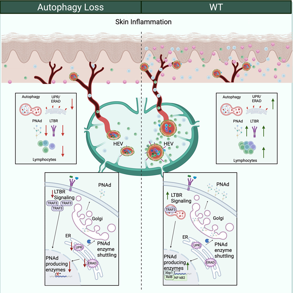
Figure 1: Graphical abstract.
The findings identify autophagy as a potential therapeutic target for diseases driven by excessive immune cell recruitment.
High endothelial venules (HEVs) are specialised blood vessels found within secondary lymphoid organs, including lymph nodes. Unlike conventional blood vessels, HEVs express high levels of peripheral node addressin (PNAd), a surface molecule that binds circulating lymphocytes and enables them to exit the bloodstream and enter lymphoid tissues where immune responses are initiated.
During inflammation, HEVs expand to accommodate increased immune cell trafficking. However, the mechanisms that preserve their specialised structure and function have remained poorly understood.
Using a combination of single-cell transcriptomics, proteomics, intravital imaging and genetic mouse models, the researchers found that HEVs exhibit substantially higher levels of autophagy than other vascular endothelial cells.
Autophagy, a cellular recycling process that removes damaged proteins and organelles, was shown to be essential for maintaining HEV identity. When autophagy was genetically disrupted, HEVs lost their specialised characteristics through a process of dedifferentiation, resulting in reduced production of PNAd and impaired recruitment of lymphocytes into lymphoid tissues.
Mechanistically, autophagy supported signalling through the lymphotoxin β receptor (LTβR) pathway while sustaining the unfolded protein response, both of which are required to maintain the specialised molecular programme that defines HEVs.
The functional consequences of disrupting autophagy were particularly evident in a mouse model of psoriasis, a chronic inflammatory skin disease driven by excessive immune activation.
Both inhibition of autophagy and blockade of LTβR signalling impaired HEV function, limiting the migration of lymphocytes into inflamed tissues. This reduction in immune cell infiltration was accompanied by decreased inflammatory cytokine production and a marked improvement in skin inflammation.
These findings suggest that HEVs actively contribute to chronic inflammatory diseases by sustaining the continuous influx of immune cells into affected tissues.
Rather than targeting immune cells directly, the study highlights the possibility of modulating the specialised blood vessels that control immune cell trafficking. By disrupting the mechanisms that preserve HEV function, it may be possible to reduce harmful immune cell infiltration while leaving broader immune function intact.
As excessive lymphocyte recruitment underlies numerous autoimmune and inflammatory disorders, targeting autophagy or LTβR signalling within HEVs could represent a novel therapeutic strategy for conditions such as psoriasis and potentially other chronic inflammatory diseases.
Journal article: Jacobs, K.A, et al. 2026. Autophagy maintains high endothelial venule identity and function during inflammation. Immunity.
Summary by Stefan Botha