


New research reveals that deadly filoviruses directly infect intestinal cells, disrupting fluid balance and driving life-threatening diarrhoea (Figure 1).
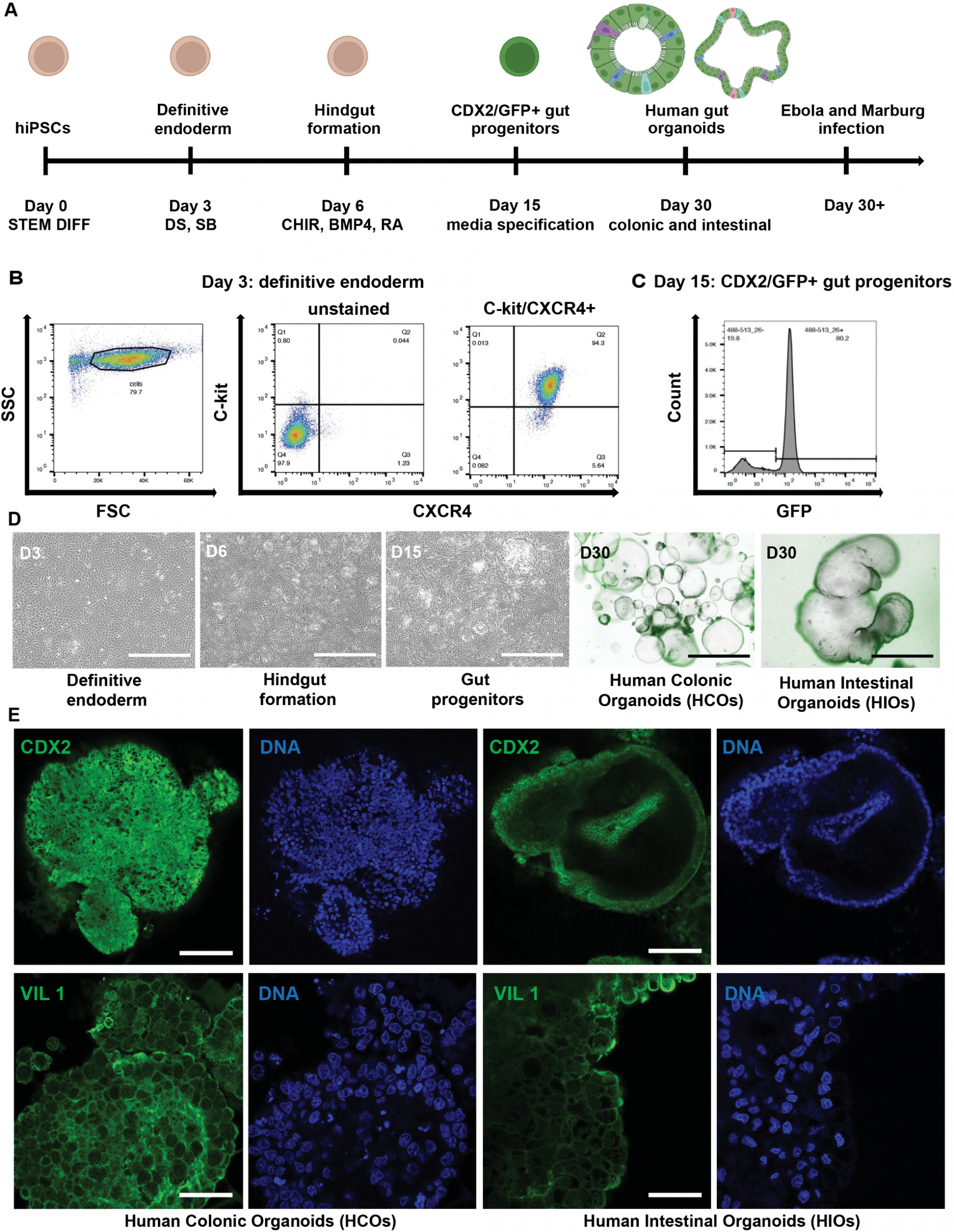
Figure 1: Directed differentiation of human iPSCs into region-specific intestinal (HIOs) and colonic (HCOs) organoids. (A) Schematic representation of the stepwise differentiation protocol used to generate gut organoids from human induced pluripotent stem cells (iPSCs). Illustrations created in BioRender. Muhlberger, E. (2025) https://BioRender.com/14baw98. (B) BU1 CDX2-GFP iPSCs were differentiated to definitive endoderm by day 3. (C) A CDX2-eGFP knock-in reporter line was used to monitor the emergence of hindgut progenitors during differentiation. (D) Gut progenitor cells were further specified into regional identities using defined media: colonic organoids (HCOs) were cultured in CKDCI media, and intestinal organoids (HIOs) were cultured in media containing CHIR, KGF, EGF, R-spondin, and Noggin. Brightfield and fluorescence imaging was performed at day 30 to visualize organoid morphology. Images were captured using a Keyence BZ-X710 fluorescence microscope. (E) Immunofluorescence staining was used to detect CDX2 and villin (VIL) (green) and Hoechst for nuclear staining (blue) in HIOs and HCOs. Confocal imaging was performed using a Zeiss LSM 710-Live Duo Confocal microscope with two-photon capability. Scale bars = 100 μm. Data shown are representative of n = 3 independent differentiations.
Ebola virus (EBOV) and Marburg virus (MARV) are among the world’s most lethal pathogens. While their devastating effects on the immune system and blood vessels are well known, one of the major causes of death, severe diarrhoea and dehydration, has remained poorly understood.
Now, a new study provides crucial insight. Published research shows that both EBOV and MARV can directly infect and replicate within human intestinal epithelial cells, fundamentally disrupting the gut’s ability to regulate fluids.
To examine what happens in the human intestine during infection, the researchers used intestinal organoids, three-dimensional “mini-guts” grown from induced pluripotent stem cells (iPSCs). These lab-grown tissues closely mimic the structure and function of the human small intestine and colon.
When the team infected these organoids with Ebola and Marburg viruses, they observed robust viral replication, confirming that the gut lining itself is a direct viral target.
By analysing gene activity in infected organoids, the researchers uncovered important differences between regions of the gut:
- Colon-like organoids showed more severe dysfunction than small-intestine-like organoids
- Key pathways controlling ion transport and fluid secretion were disrupted
- Structural damage occurred at the apical surface and cell–cell junctions, which normally regulate what passes through the intestinal wall
Together, these changes would allow uncontrolled fluid loss into the gut, mirroring the profuse, watery diarrhoea seen in patients with severe Ebola or Marburg disease.
The study also found that infected gut tissues mounted a delayed innate immune response, particularly in the activation of interferon-stimulated genes, which are normally critical for early antiviral defence. This delay may give the virus time to replicate unchecked, compounding tissue damage before immune defences engage.
By directly targeting the intestinal lining, the viruses disrupt both barrier integrity and fluid regulation, leading to rapid dehydration even when supportive care is available.
Understanding these mechanisms opens new avenues for:
- Targeted supportive therapies to preserve gut barrier function
- Improved clinical management of fluid loss
- Antiviral strategies aimed at protecting epithelial tissues, not just immune cells
By combining advanced stem-cell technology with high-containment virology, this work provides one of the most detailed pictures yet of how filoviruses damage human organs at the cellular level without requiring invasive patient sampling.
Ebola and Marburg viruses don’t just trigger systemic illness they directly infect and disable the gut lining. By disrupting fluid transport and delaying immune responses, these viruses drive the catastrophic diarrhea that makes filovirus infections so deadly. This new insight could guide better treatments to save lives during future outbreaks.
Journal article: Flores, E. Y., et al. 2025. Filovirus Infection disrupts epithelial barrier function and ion transport in human iPSC-derived gut organoids. PLoS Pathogens.
Summary by Stefan Botha


