


Chikungunya virus infections are often acute, but for 30% to 60% of patients, especially women, the virus leaves a troubling legacy: chronic, severe joint pain that can persist for years. This long-term condition mimics rheumatoid arthritis in both symptoms and immune profile, leading scientists to investigate whether T cells are to blame for the lingering inflammation.
A new study provides the first detailed map of how the human immune system, specifically T cells, recognizes and responds to the Chikungunya virus, a mosquito-borne pathogen known for causing long-lasting joint pain (Figure 1). This breakthrough brings researchers closer to developing T cell–based vaccines and therapies that could precisely target the virus and potentially prevent chronic symptoms.
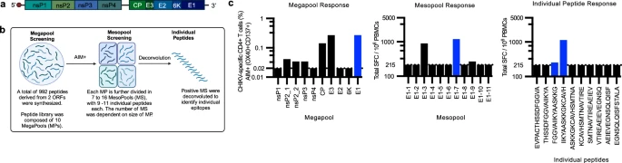
Figure 1: Experimental workflow for screening CD4+ T cell epitopes in CHIKV. a Schematic representation of CHIKV proteome comprising four non-structural (nsP1, nsP2, nsP3 and nsP4) and five structural proteins (Capsid or CP, E3, E2 and E1). b Workflow of epitope screening. All donors were tested in the AIM assay by stimulation with 10 megapools (MP) corresponding to each CHIKV protein (nsP1, nsP2_1, nsP2_2, nsP3, nsP4, CP, E3, E2, 6 K, E1). Positive donors in the AIM assay (OX40+ CD137+ or OX40+ CD40L+ ) were tested in the FluoroSpot assay by stimulating with smaller pools of MP, called mesopools (MS), each of which contained 9–11 individual peptides. Each MS was deconvoluted to determine individual epitopes. Created in BioRender. Weiskopf, D. (2025) https://BioRender.com/2bdm25t. c An example of the experimental workflow for the responses of one donor to the E1 protein. The first panel shows responses in the AIM assay to all CHIKV MPs tested. The middle panel shows SFCs per million PBMCs to each MS of the E1 protein. The third panel depicts responses to individual peptides in E1-7 MS. The dotted line indicates the threshold of positivity. The blue highlighted bars depict an example of a positive response from one donor.
Previous work showed that people with chronic Chikungunya disease carry inflammatory CD4+ T cells with a profile similar to those found in autoimmune disorders. The new study expands on that work by mapping out which viral components (epitopes) these T cells respond to.
To identify the immunodominant parts of the virus, the region’s most frequently recognized by T cells, the researchers used a novel method. They broke the virus into small protein fragments (peptides) and exposed immune cells from infected individuals to these peptides to see which ones triggered the strongest T cell responses.
The result: a comprehensive atlas of T cell epitopes for Chikungunya virus. This map serves as a valuable tool for guiding vaccine design and pinpointing therapeutic targets that could modulate immune responses more precisely. They analysed CD4+ T cells from two groups of people: those who developed chronic joint pain after infection and those who cleared the virus without lasting symptoms.
Surprisingly, both groups had T cells that targeted the same viral epitopes. This means that chronic disease isn’t caused by the immune system recognizing different parts of the virus. Instead, the issue may lie in how long these T cells persist and where the virus might be hiding in the body to keep triggering them..
With Chikungunya virus spreading to new geographic regions, thanks in part to expanding mosquito habitats, understanding its long-term effects is becoming increasingly urgent.
Journal article: Agarwal, R., et al,. 2025. Identification of immunogenic and cross-reactive chikungunya virus epitopes for CD4+ T cells in chronic chikungunya disease, Nature Communications.
Summary by Stefan Botha


