


Feline heartworm disease, caused by the parasite Dirofilaria immitis, presents diagnostic challenges, especially in early infections with immature worms. This study assessed thoracic radiographic changes in cats from an endemic area to identify diagnostic markers of heartworm-associated respiratory disease (HARD). A total of 123 cats were examined, divided into three groups: seronegative, asymptomatic seropositive, and seropositive with clinical signs. Radiographic measurements focused on key vascular ratios (CrPA/R4, CdPA/R9), comparing pulmonary artery diameters with rib diameters to assess arterial enlargement.
Further reading: Hematocrit Fluctuations in HIV Patients Co-infected with Malaria Parasites: A Comprehensive Review
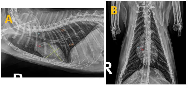
Figure 1 Thoracic radiographs of a cat seropositive for anti-Dirofilaria-immitis antibodies and presenting with clinical signs. The measurements taken during this study are shown as follows: (A) Right laterolateral projection illustrating the measurements of the caudal vena cava (CVC) and aorta (Ao) (in orange), the fourth rib (R4) (in blue), the right cranial pulmonary artery (CrPA) (in red) and the vertebral heart score (in green). (B) Dorsoventral projection displaying the measurement of the right caudal pulmonary artery (CdPA) (in red) in relation to the ninth rib (R9) (in blue).
The results demonstrated significant differences in the CrPA/R4 and CdPA/R9 ratios between infected and healthy cats, indicating vascular changes that can signal heartworm infection even in asymptomatic animals. Cats with clinical signs displayed marked bronchointerstitial patterns in 62.8% of cases, highlighting parenchymal abnormalities typically associated with advanced infection stages. However, the vertebral heart score (VHS) did not significantly differ across groups, suggesting that while some cardiac enlargement may appear, VHS is not a reliable indicator of HARD in cats.
These findings confirm that radiographic evaluations, particularly vascular measurements, provide a non-invasive method for detecting early arterial changes in cats with D. immitis infection. Despite this, some infected cats showed normal radiographs, emphasizing that radiographic assessments alone may not capture all cases. Thus, combining radiographic measurements with serological testing improves diagnostic accuracy and supports early detection of HARD, which is especially crucial in cases where infections have not yet produced clinical signs.
This integrative diagnostic approach enhances the ability to detect heartworm-associated abnormalities in feline patients, assisting veterinarians in identifying early signs of HARD. The study underscores the importance of quantifiable radiographic indices and combined diagnostic methods for effective management and intervention in endemic areas.
Journal article: Falcón-Cordón, Soraya, et al. “Radiological Evaluation of Vascular Structures in Cats Infected with Immature Worms of Dirofilaria Immitis.” Animals.
Summary by Faith Oluwamakinde


