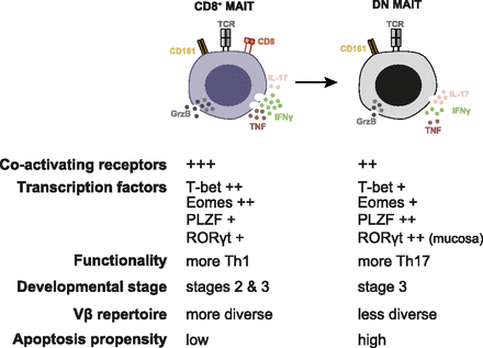
Properties and relationship of the main MAIT cell subsets. (Source: Dias et al., 2018 PNAS)
Mucosal associated invariant T (MAIT) cells are MHC class I-like restricted T cells, that recognise bacterial riboflavin metabolites. Studies have shown that MAIT cells can be activated either a MR1-dependent manner or MR1-independent manner via IL-12/IL-18 activation. Upon activation they can produce pro-inflammatory cytokines (IFN-𝛄, IL-17) and cytotoxic molecules. In adulthood circulating (non-thymus) MAIT cells predominantly express CD8⍺, however a subset of CD4-CD8- (double negative, DN) MAIT cells have also be detected and characterised. Whether these two phenotypes represents truly distinct functional MAIT subsets is unclear.
Study by Dias et al., aimed to determine the difference in transcriptional, activation and functional capacity of CD8+ and DN MAIT cells. They defined MAIT cells as CD161hiTRAV-1-2+ MR1-tetramer+ T cells. CD8+ MAIT cells expressed higher levels of activation markers (CD9, CD27, NKG2D, PD-1), functional (IFN-𝛄, TNF) and cytotoxic markers (granzyme B, granulysin, perforin) than DN MAIT cells. Suggesting that CD8+ MAIT cells are more responsive and functional than DN cells, this was further demonstrated by increased expression of T-bet, Eomes and up-regulation of pro-inflammatory (mRNA) transcripts in CD8+ MAIT cells than DN MAIT cells. Surprisingly, DN MAIT cells detected in tissue expressed higher levels of PLZF, ROR𝛄t and Helios than CD8+ MAIT cells, however this did not translate to significantly higher levels of IL-17 production compared with CD8+ MAIT cells.
How do DN MAIT cells form?
Three stages of MAIT cell development in the thymus have been characterised. DN MAIT cells first appear in the 3rd stage of MAIT cell development, however at very low frequencies and are observed at significantly more abundant levels in circulation (blood). Upon in vitro activation by antigen and in long term cell culture assays CD8+ MAIT cells down regulated expression of CD8 resulting in increased proportion of DN MAIT cells. This down regulation occurred in an MR-1 dependent manner, suggesting that a proportion of DN MAIT cells differentiate from CD8+ MAIT cells.
In summary findings by Dias et al., defined CD8+ and DN MAIT cells as two distinct populations, where DN MAIT cells are more differentiated and less functional than CD8+ MAIT cells. They also showed that DN MAIT cells can be derived from CD8+ MAIT cells, elucidating why they are observed at increased proportions in circulation during adulthood than during thymic development.
Journal Article: Dias et al., 2018., The CD4−CD8− MAIT cell subpopulation is a functionally distinct subset developmentally related to the main CD8+ MAIT cell pool. PNAS
Article by Cheleka AM Mpande