


Differentiating tuberculous pleural effusion (TPE) from para-pneumonic pleural effusion (PPE) is challenging due to overlapping clinical signs. Conventional diagnostic methods, such as adenosine deaminase levels, can misclassify cases. This study investigates myeloid-derived suppressor cells (MDSCs) as a potential marker for early discrimination between TPE and PPE.
A prospective cohort of 47 patients with pleural effusion was analysed at Seoul National University Bundang Hospital. Researchers used flow cytometry to quantify polymorphonuclear-MDSCs (PMN-MDSCs) and other immune cell populations in pleural fluid samples. Diagnostic accuracy was assessed using the receiver operating characteristic (ROC) curve.
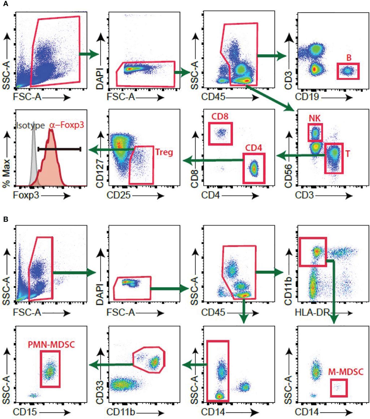
Figure 1: Gating strategy of immune cells. (A) B, NK, T, Treg cells (B) PMN-MDSC, M-MDSC.
PMN-MDSCs were significantly higher in PPE than in TPE cases, suggesting their diagnostic potential. ROC analysis indicated PMN-MDSCs provided a high area under the curve (AUC), outperforming traditional markers. Additionally, PMN-MDSCs elevated reactive oxygen species (ROS) levels and suppressed T-cell interferon-gamma production, contributing to the immune profile difference between TPE and PPE.
PMN-MDSCs are a promising marker for distinguishing TPE from PPE, potentially enabling faster and more accurate diagnosis. Integrating MDSC analysis into clinical practice could improve treatment decisions and patient outcomes in regions with high tuberculosis prevalence.
Summary by Faith Oluwamakinde


