


For almost 50 years, scientists have known that Plasmodium parasites invade red blood cells using a specialised ring-shaped structure called the moving junction. Although this structure is essential for infection, its precise role has remained elusive because it forms and disappears within less than a minute during parasite invasion.
In a recent study, researchers have captured the moving junction in unprecedented detail, revealing that it is not simply a passive gateway as previously believed (Figure 1). Instead, the study, shows that the moving junction functions as an active molecular machine that remodels the host cell membrane, helping the parasite force its way into red blood cells.
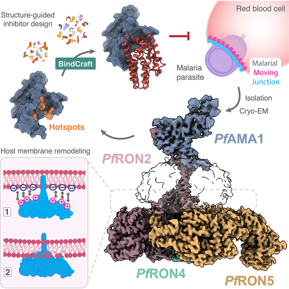
Figure 1: Graphical abstract.
To study this highly transient structure, the researchers chemically stalled malaria parasites now they began invading red blood cells. This allowed them to isolate the intact moving junction and visualise it using cryo-electron microscopy, producing the first high-resolution three-dimensional structure of its core protein complex.
The complex consists of four parasite proteins, AMA1, RON2, RON4 and RON5, which together form the structural foundation of the moving junction. Rather than resembling a simple ring, the complex adopts a distinctive “sailboat-like” architecture, with AMA1 forming the “sail” above the cell surface while the RON proteins create a broad “hull” positioned against the host cell membrane.
The structural analysis revealed features typically found in proteins that bend and reshape biological membranes. The underside of the complex contains positively charged regions that interact with the host membrane, alongside short helical segments that insert into the membrane like wedges.
To confirm their findings, the team synthesised these wedge-like helices and tested them on artificial membrane vesicles. The helices disrupted and thinned the membranes, whereas modified, weakened versions had little effect. These experiments suggest that the moving junction actively deforms the host cell membrane, working together with the parasite’s internal motor to facilitate entry into red blood cells.
This discovery challenges the long-standing view that the moving junction serves only as a static anchor through which the parasite pulls itself during invasion.
Malaria continues to cause approximately 600,000 deaths each year, predominantly among young children in sub-Saharan Africa, while resistance to existing antimalarial drugs continues to rise. Because every malaria infection depends on successful invasion of red blood cells, the moving junction has long been considered an attractive therapeutic target.
Using their newly determined structure, the researchers applied machine learning-assisted protein design to create a small synthetic protein that disrupts the interaction between AMA1 and its binding partner, preventing assembly of the moving junction. In laboratory experiments, the designed mini-protein blocked parasite invasion of red blood cells in a dose-dependent manner without affecting cells that were already infected, indicating that it specifically targets the invasion process.
Although this engineered protein is an early proof of concept rather than a clinical drug candidate, the work demonstrates how high-resolution structural biology can guide the rational design of next-generation antimalarial therapeutics. The findings may also inform future malaria vaccine development by providing new insights into how protective antibodies target the invasion machinery.
Journal article: Haile, M.T., et al. 2026. Structural basis for host membrane binding and remodeling by invading malaria parasites. Cell.
Summary by Stefan Botha