


The thymus is often described as the training ground of the immune system, where developing T cells are selected and shaped before entering the body. New research has revealed that this process is far more specialised than previously appreciated, uncovering how a unique population of immune cells, mucosal-associated invariant T (MAIT) cells, is programmed in the thymus to protect tissues throughout the body (Figure 1).
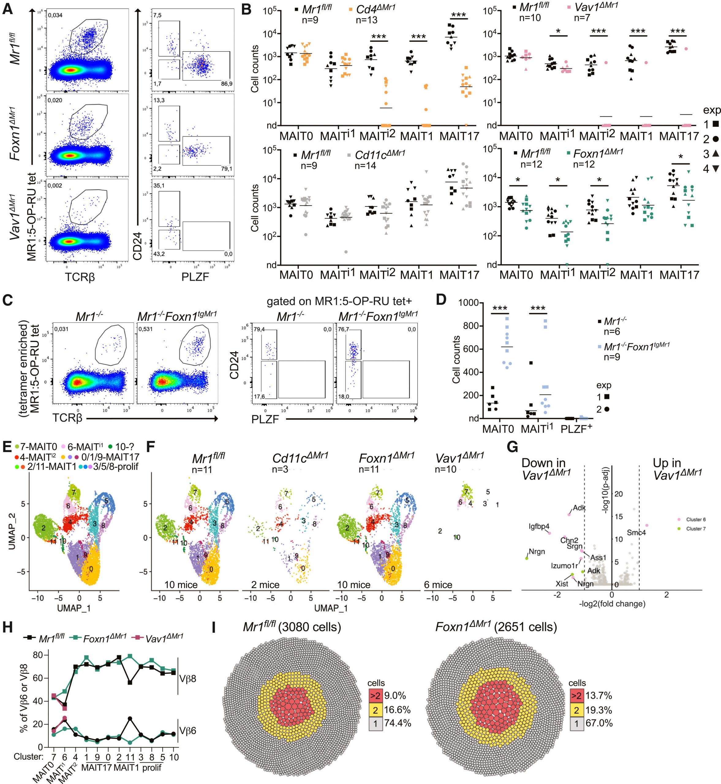
Figure 1: MR1 expression by TECs and DP thymocytes regulates thymic MAIT cell development.
The study reveals that MAIT cells undergo distinct developmental pathways compared with another related immune population, invariant natural killer T (iNKT) cells). The findings show that thymic signals determine where MAIT cells migrate, how they specialise, and how they contribute to tissue repair during inflammation.
MAIT and iNKT cells belong to a family of innate-like T cells that combine features of innate and adaptive immunity. Unlike conventional T cells, which recognise diverse peptide antigens presented by highly variable MHC molecules, these cells recognise conserved molecular signals presented by specialised antigen-presenting molecules.
MAIT cells detect microbial metabolites derived from the vitamin B2 biosynthesis pathway through the molecule MR1, while iNKT cells recognise lipid antigens presented by CD1d. This allows them to rapidly respond to infections, tissue injury, and inflammatory signals without requiring the same activation process as conventional T cells.
Both MAIT and iNKT cells emerge in the thymus with an activated memory-like phenotype, allowing them to rapidly produce inflammatory cytokines and participate in immune defence. However, despite sharing many developmental features, these two cell populations have different distributions throughout the body, suggesting that their development is controlled by distinct mechanisms.
The researchers discovered that thymic epithelial cells (TECs) play a previously unrecognised role in MAIT cell development. Expression of MR1 by thymic epithelial cells helps select developing MAIT cells, allowing them to mature appropriately.
In contrast, iNKT cell development was unaffected by CD1d expression on thymic epithelial cells, indicating that iNKT selection depends primarily on interactions with developing double-positive thymocytes.
This distinction reveals that, although MAIT and iNKT cells follow similar developmental programs, their earliest stages are shaped by different cellular environments within the thymus.
The study also challenges the traditional view that MAIT and iNKT cells leave the thymus only after reaching full maturity. Instead, researchers found that MAIT cells exit the thymus at multiple developmental stages, with different subsets carrying distinct instructions for where they will migrate.
One important discovery was the role of chemokine receptors in directing tissue-specific migration. Semi-mature MAIT cells expressing CCR9 preferentially migrated to the intestine, where they could undergo further maturation after encountering signals from the local environment.
This suggests that the thymus does not simply produce mature immune cells, it generates populations with pre-programmed tissue preferences that allow them to rapidly populate specific organs.
The researchers further demonstrated that thymic output continues to contribute to MAIT and iNKT cell populations throughout adulthood. This challenges the idea that these cells are generated only during early life and then maintained independently.
Instead, ongoing production from the thymus helps maintain immune surveillance and tissue homeostasis. The importance of this pathway became particularly evident during intestinal inflammation. In a model of colitis, increased production of MAIT cell ligands in peripheral tissues triggered expansion and activation of MAIT17 cells, a specialised subset that produces IL-17 and contributes to epithelial repair.
These activated thymic-derived MAIT17 cells migrated to the colon, suggesting the existence of a feedback mechanism: tissue damage increases signals recognised by MAIT cells, which then promotes production and recruitment of repair-oriented MAIT populations.
This finding highlights the dual role of MAIT cells in immunity. While they can contribute to antimicrobial defence, they also act as regulators of tissue integrity by promoting repair responses after injury.
By revealing how the thymus prepares MAIT cells for specialised roles across different organs, this work expands our understanding of immune cell development and highlights the thymus as a key regulator of lifelong tissue immunity.
Journal article: Paiva, R.A., et al. 2026. MAIT cells egress the thymus at several maturation stages with selective capacity to seed different tissues. Immunity.
Summary by Stefan Botha


